1K9G | pdb_00001k9g

Crystal Structure of the Complex of Cryptolepine-d(CCTAGG)2


Experimental Data Snapshot

  • Method: X-RAY DIFFRACTION
  • Resolution: 1.40 Å
  • R-Value Free: 
    0.227 (Depositor) 
  • R-Value Work: 
    0.208 (Depositor) 
  • R-Value Observed: 
    0.209 (Depositor) 

wwPDB Validation 3D Report Full Report

Validation slider image for 1K9G

This is version 1.3 of the entry. See complete history

Literature

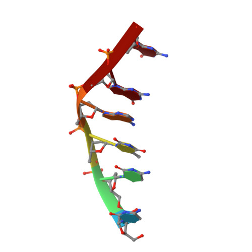
The antimalarial and cytotoxic drug cryptolepine intercalates into DNA at cytosine-cytosine sites.

Lisgarten, J.N.Coll, M.Portugal, J.Wright, C.W.Aymami, J.

(2002) Nat Struct Biol 9: 57-60

  • DOI: https://doi.org/10.1038/nsb729
  • Primary Citation Related Structures: 
    1K9G

  • PubMed Abstract: 

    Cryptolepine, a naturally occurring indoloquinoline alkaloid used as an antimalarial drug in Central and Western Africa, has been found to bind to DNA in a formerly unknown intercalation mode. Evidence from competition dialysis assays demonstrates that cryptolepine is able to bind CG-rich sequences containing nonalternating CC sites. Here we show that cryptolepine interacts with the CC sites of the DNA fragment d(CCTAGG)(2) in a base-stacking intercalation mode. This is the first DNA intercalator complex, from approximately 90 solved by X-ray crystallography, to bind a nonalternating (pyrimidine-pyrimidine) DNA sequence. The asymmetry of the drug induces a perfect stacking with the asymmetric site, allowing for the stability of the complex in the absence of hydrogen bonding interactions. The crystal structure of this antimalarial drug-DNA complex provides evidence for the first nonalternating intercalation and, as such, provides a basis for the design of new anticancer or antimalarial drugs.


  • Organizational Affiliation
    • Institut de Biologia Molecular de Barcelona, C.S.I.C., Jordi Girona 18, 08034 Barcelona, Spain.

Macromolecule Content 

  • Total Structure Weight: 2.27 kDa 
  • Atom Count: 193 
  • Modeled Residue Count: 6 
  • Deposited Residue Count: 6 
  • Unique nucleic acid chains: 1

Macromolecules

Find similar nucleic acids by:  Sequence
Entity ID: 1
MoleculeChains LengthOrganismImage
5'-D(*CP*CP*TP*AP*GP*G)-3'6N/A
Sequence Annotations
Expand
Reference Sequence

Small Molecules

Ligands 1 Unique
IDChains Name / Formula / InChI Key2D Diagram3D Interactions
DR1

Query on DR1


Download:Ideal Coordinates CCD File
B [auth A],
C [auth A]
5-METHYL-5H-INDOLO[3,2-B]QUINOLINE
C16 H12 N2
KURWKDDWCJELSV-UHFFFAOYSA-N

Experimental Data & Validation

Experimental Data

  • Method: X-RAY DIFFRACTION
  • Resolution: 1.40 Å
  • R-Value Free:  0.227 (Depositor) 
  • R-Value Work:  0.208 (Depositor) 
  • R-Value Observed: 0.209 (Depositor) 
Space Group: P 64
Unit Cell:
Length ( Å )Angle ( ˚ )
a = 29.96α = 90
b = 29.96β = 90
c = 39.65γ = 120
Software Package:
Software NamePurpose
AMoREphasing
SHELXL-97refinement
MAR345data collection
SCALEPACKdata scaling

Structure Validation

View Full Validation Report


Entry History 

Deposition Data

Revision History  (Full details and data files)

  • Version 1.0: 2001-11-30
    Type: Initial release
  • Version 1.1: 2008-04-27
    Changes: Version format compliance
  • Version 1.2: 2011-07-13
    Changes: Version format compliance
  • Version 1.3: 2024-02-07
    Changes: Data collection, Database references, Derived calculations, Refinement description