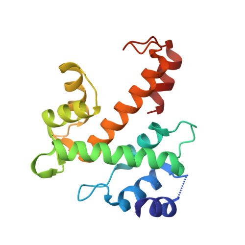
Structure of Ca(2+)-loaded human grancalcin.
Jia, J., Borregaard, N., Lollike, K., Cygler, M.(2001) Acta Crystallogr D Biol Crystallogr 57: 1843-1849
- PubMed: 11717497 Search on PubMed
- DOI: https://doi.org/10.1107/s0907444901016511
- Primary Citation Related Structures:
1K94, 1K95 - PubMed Abstract:
Grancalcin is a cytosolic Ca(2+)-binding protein originally identified in human neutrophils. It belongs to a new class of EF-hand proteins, called PEF proteins, which contain five EF-hand motifs. At the N-terminus of grancalcin there is a approximately 50 residue-long segment rich in glycines and prolines. The fifth EF-hand, unpaired within the monomer, provides a means for dimerization through pairing with its counterpart in a second molecule. The structure of full-length grancalcin in the apo form and with one EF3 within the dimer occupied by a Ca(2+) ion have been determined. Although the N-terminal segment was present in the molecule, this part was disordered in the crystals. Here, the structure of a truncated form of grancalcin, which is lacking 52 N-terminal residues, in the presence and absence of Ca(2+) is presented. In the Ca(2+)-bound form the ions are found in the EF1 and EF3 hands. Binding of Ca(2+) to these two EF hands produces only minor conformational changes, mostly within the EF1 Ca(2+)-binding loop. This observation supports the hypothesis, formulated on the basis of the structure of a homologous protein ALG-2 which shows significant differences in the orientation of EF4 and EF5 compared with grancalcin, that calcium is a necessary factor but not sufficient alone for inducing a significant conformational change in PEF proteins.
- Biotechnology Research Institute, National Research Council of Canada, 6100 Royalmount Avenue, Montreal, Quebec H4P 2R2, Canada.
Organizational Affiliation: