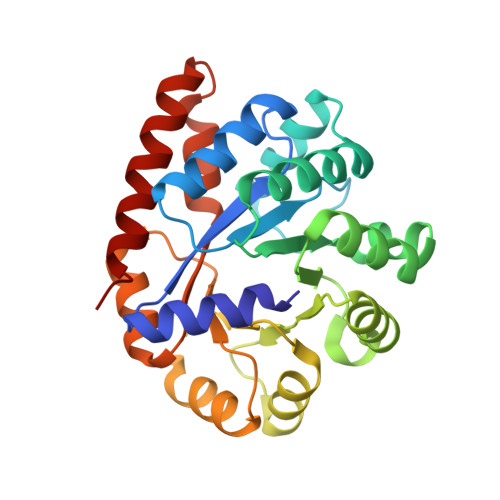
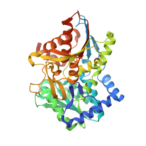
1K8Y

CRYSTAL STRUCTURE OF THE TRYPTOPHAN SYNTHASE BETA-SER178PRO MUTANT COMPLEXED WITH D,L-ALPHA-GLYCEROL-3-PHOSPHATE


Experimental Data Snapshot

  • Method: X-RAY DIFFRACTION
  • Resolution: 1.50 Å
  • R-Value Free: 0.209 
  • R-Value Work: 0.171 
  • R-Value Observed: 0.173 

Starting Model: experimental
View more details

wwPDB Validation   3D Report Full Report


Ligand Structure Quality Assessment 


This is version 1.4 of the entry. See complete history


Literature

Crystal structure of the beta Ser178--> Pro mutant of tryptophan synthase. A "knock-out" allosteric enzyme.

Weyand, M.Schlichting, I.Herde, P.Marabotti, A.Mozzarelli, A.

(2002) J Biol Chem 277: 10653-10660

  • DOI: https://doi.org/10.1074/jbc.M111031200
  • Primary Citation of Related Structures:  
    1K7X, 1K8Y, 1K8Z

  • PubMed Abstract: 

    The catalytic activity of the pyridoxal 5'-phosphate-dependent tryptophan synthase alpha(2)beta(2) complex is allosterically regulated. The hydrogen bond between the helix betaH6 residue betaSer(178) and the loop alphaL6 residue Gly(181) was shown to be critical in ligand-induced intersubunit signaling, with the alpha-beta communication being completely lost in the mutant betaSer(178) --> Pro (Marabotti, A., De Biase, D., Tramonti, A., Bettati, S., and Mozzarelli, A. (2001) J. Biol. Chem. 276, 17747-17753). The structural basis of the impaired allosteric regulation was investigated by determining the crystal structures of the mutant betaSer(178) --> Pro in the absence and presence of the alpha-subunit ligands indole-3-acetylglycine and glycerol 3-phosphate. The mutation causes local and distant conformational changes especially in the beta-subunit. The ligand-free structure exhibits larger differences at the N-terminal part of helix betaH6, whereas the enzyme ligand complexes show differences at the C-terminal side. In contrast to the wild-type enzyme loop alphaL6 remains in an open conformation even in the presence of alpha-ligands. This effects the equilibrium between active and inactive conformations of the alpha-active site, altering k(cat) and K(m), and forms the structural basis for the missing allosteric communication between the alpha- and beta-subunits.


  • Organizational Affiliation

    Max-Planck-Institut für Molekulare Physiologie, Abteilung für Physikalische Biochemie, D-44227 Dortmund, Germany.


Macromolecules
Find similar proteins by:  (by identity cutoff)  |  3D Structure
Entity ID: 1
MoleculeChains Sequence LengthOrganismDetailsImage
TRYPTOPHAN SYNTHASE ALPHA CHAIN268Salmonella enterica subsp. enterica serovar TyphimuriumMutation(s): 0 
Gene Names: TRPA/TRPB
EC: 4.2.1.20
UniProt
Find proteins for P00929 (Salmonella typhimurium (strain LT2 / SGSC1412 / ATCC 700720))
Explore P00929 
Go to UniProtKB:  P00929
Entity Groups  
Sequence Clusters30% Identity50% Identity70% Identity90% Identity95% Identity100% Identity
UniProt GroupP00929
Sequence Annotations
Expand
  • Reference Sequence
Find similar proteins by:  (by identity cutoff)  |  3D Structure
Entity ID: 2
MoleculeChains Sequence LengthOrganismDetailsImage
TRYPTOPHAN SYNTHASE BETA CHAIN396Salmonella enterica subsp. enterica serovar TyphimuriumMutation(s): 1 
Gene Names: TRPA/TRPB
EC: 4.2.1.20
UniProt
Find proteins for P0A2K1 (Salmonella typhimurium (strain LT2 / SGSC1412 / ATCC 700720))
Explore P0A2K1 
Go to UniProtKB:  P0A2K1
Entity Groups  
Sequence Clusters30% Identity50% Identity70% Identity90% Identity95% Identity100% Identity
UniProt GroupP0A2K1
Sequence Annotations
Expand
  • Reference Sequence
Experimental Data & Validation

Experimental Data

  • Method: X-RAY DIFFRACTION
  • Resolution: 1.50 Å
  • R-Value Free: 0.209 
  • R-Value Work: 0.171 
  • R-Value Observed: 0.173 
  • Space Group: C 1 2 1
Unit Cell:
Length ( Å )Angle ( ˚ )
a = 183.142α = 90
b = 60.465β = 94.63
c = 67.44γ = 90
Software Package:
Software NamePurpose
CNSrefinement
REFMACrefinement
XDSdata reduction
XSCALEdata scaling
CNSphasing

Structure Validation

View Full Validation Report


Ligand Structure Quality Assessment 


Entry History 

Deposition Data

Revision History  (Full details and data files)

  • Version 1.0: 2002-06-19
    Type: Initial release
  • Version 1.1: 2008-04-27
    Changes: Version format compliance
  • Version 1.2: 2011-07-13
    Changes: Non-polymer description, Version format compliance
  • Version 1.3: 2021-10-27
    Changes: Database references, Derived calculations
  • Version 1.4: 2023-08-16
    Changes: Data collection, Refinement description