1K8W

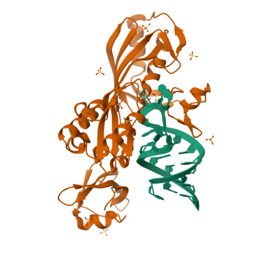
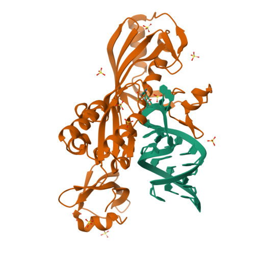
Crystal structure of the E. coli pseudouridine synthase TruB bound to a T stem-loop RNA


Experimental Data Snapshot

  • Method: X-RAY DIFFRACTION
  • Resolution: 1.85 Å
  • R-Value Free: 0.211 
  • R-Value Work: 0.184 
  • R-Value Observed: 0.184 

wwPDB Validation   3D Report Full Report


This is version 1.3 of the entry. See complete history


Literature

Cocrystal structure of a tRNA Psi55 pseudouridine synthase: nucleotide flipping by an RNA-modifying enzyme.

Hoang, C.Ferre-D'Amare, A.R.

(2001) Cell 107: 929-939

  • DOI: https://doi.org/10.1016/s0092-8674(01)00618-3
  • Primary Citation of Related Structures:  
    1K8W

  • PubMed Abstract: 

    Pseudouridine (Psi) synthases catalyze the isomerization of specific uridines in cellular RNAs to pseudouridines and may function as RNA chaperones. TruB is responsible for the Psi residue present in the T loops of virtually all tRNAs. The close homolog Cbf5/dyskerin is the catalytic subunit of box H/ACA snoRNPs. These carry out the pseudouridylation of eukaryotic rRNA and snRNAs. The 1.85 A resolution structure of TruB bound to RNA reveals that this enzyme recognizes the preformed three-dimensional structure of the T loop, primarily through shape complementarity. It accesses its substrate uridyl residue by flipping out the nucleotide and disrupts the tertiary structure of tRNA. Structural comparisons with TruB demonstrate that all Psi synthases are descended from a common molecular ancestor.


  • Organizational Affiliation

    Division of Basic Sciences, Fred Hutchinson Cancer Research Center, 1100 Fairview Avenue North, Seattle, WA 98109, USA.


Macromolecules

Find similar proteins by:  (by identity cutoff)  |  3D Structure
Entity ID: 2
MoleculeChains Sequence LengthOrganismDetailsImage
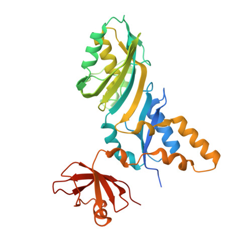
tRNA Pseudouridine Synthase BB [auth A]327Escherichia coliMutation(s): 0 
EC: 4.2.1.70 (PDB Primary Data), 5.4.99.25 (UniProt)
UniProt
Find proteins for P60340 (Escherichia coli (strain K12))
Explore P60340 
Go to UniProtKB:  P60340
Entity Groups  
Sequence Clusters30% Identity50% Identity70% Identity90% Identity95% Identity100% Identity
UniProt GroupP60340
Sequence Annotations
Expand
  • Reference Sequence

Find similar nucleic acids by:  Sequence   |   3D Structure  

Entity ID: 1
MoleculeChains LengthOrganismImage
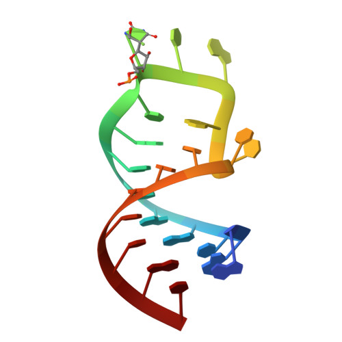
5'-R(*GP*GP*CP*AP*AP*CP*GP*GP*UP*(FHU)P*CP*GP*AP*UP*CP*CP*CP*GP*UP*UP*GP*C)-3'A [auth B]22N/A
Sequence Annotations
Expand
  • Reference Sequence
Small Molecules
Ligands 1 Unique
IDChains Name / Formula / InChI Key2D Diagram3D Interactions
SO4
Query on SO4

Download Ideal Coordinates CCD File 
C [auth B]
D [auth A]
E [auth A]
F [auth A]
G [auth A]
C [auth B],
D [auth A],
E [auth A],
F [auth A],
G [auth A],
H [auth A],
I [auth A],
J [auth A],
K [auth A],
L [auth A]
SULFATE ION
O4 S
QAOWNCQODCNURD-UHFFFAOYSA-L
Experimental Data & Validation

Experimental Data

  • Method: X-RAY DIFFRACTION
  • Resolution: 1.85 Å
  • R-Value Free: 0.211 
  • R-Value Work: 0.184 
  • R-Value Observed: 0.184 
  • Space Group: C 1 2 1
Unit Cell:
Length ( Å )Angle ( ˚ )
a = 145.054α = 90
b = 40.361β = 110.6
c = 77.987γ = 90
Software Package:
Software NamePurpose
CNSrefinement
DENZOdata reduction
SCALEPACKdata scaling
CNSphasing

Structure Validation

View Full Validation Report


Entry History 

Deposition Data

Revision History  (Full details and data files)

  • Version 1.0: 2001-12-31
    Type: Initial release
  • Version 1.1: 2008-04-27
    Changes: Version format compliance
  • Version 1.2: 2011-07-13
    Changes: Version format compliance
  • Version 1.3: 2024-02-07
    Changes: Data collection, Database references, Derived calculations