1K66 | pdb_00001k66

Crystal Structure of the Cyanobacterial Phytochrome Response Regulator, RcpB


Experimental Data Snapshot

  • Method: X-RAY DIFFRACTION
  • Resolution: 1.75 Å
  • R-Value Free: 
    0.250 (Depositor), 0.249 (DCC) 
  • R-Value Work: 
    0.216 (Depositor), 0.215 (DCC) 
  • R-Value Observed: 
    0.216 (Depositor) 

wwPDB Validation 3D Report Full Report

Validation slider image for 1K66

This is version 1.3 of the entry. See complete history

Literature

Crystal structures of two cyanobacterial response regulators in apo- and phosphorylated form reveal a novel dimerization motif of phytochrome-associated response regulators

Benda, C.Scheufler, C.Tandeau de Marsac, N.Gaertner, W.

(2004) Biophys J 87: 476-487

  • DOI: https://doi.org/10.1529/biophysj.103.033696
  • Primary Citation Related Structures: 
    1K66, 1K68

  • PubMed Abstract: 

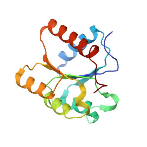
    The structures of two response regulators (RRs) from the cyanobacterium Calothrix PCC7601, RcpA and RcpB, were solved to 1.9- and 1.75-A resolution, respectively. RcpA was found in phosphorylated and RcpB in nonphosphorylated form. Both RRs are members of phytochrome-associated, light-sensing two-component signal transduction pathways, based on histidine kinase-mediated receptor autophosphorylation and phosphorelay to a RR. Despite the overall folding similarity to CheY-type RRs ((beta/alpha)(5)-motif), RcpA and RcpB form homodimers, irrespective of their phosphorylation state, giving insight into a signal transduction putatively different from that of other known RRs. Dimerization is accomplished by a C-terminal extension of the RR polypeptide chain, and the surface formed by H4, beta 5, and H5, which constitute a hydrophobic contact area with distinct interactions between residues of either subunit. Sequence alignments reveal that the identified dimerization motif is archetypal for phytochrome-associated RRs, making them a novel subgroup of CheY-type RRs. The protein structures of RcpA and RcpB are compared to the recently presented protein structure of Rcp1 from Synechocystis.


  • Organizational Affiliation
    • Max-Planck-Institut für Biochemie, D-82152 Martinsried, Germany.

Macromolecule Content 

  • Total Structure Weight: 33.67 kDa 
  • Atom Count: 2,750 
  • Modeled Residue Count: 298 
  • Deposited Residue Count: 298 
  • Unique protein chains: 1

Macromolecules

Find similar proteins by:|  3D Structure
Entity ID: 1
MoleculeChains  Sequence LengthOrganismDetailsImage
Phytochrome Response Regulator RcpB
A, B
149Tolypothrix sp. PCC 7601Mutation(s): 0 
Gene Names: AF309560
UniProt
Find proteins for Q8RTM8 (Tolypothrix sp. PCC 7601)
Explore Q8RTM8 
Go to UniProtKB:  Q8RTM8
Entity Groups
Sequence Clusters30% Identity50% Identity70% Identity90% Identity95% Identity100% Identity
UniProt GroupQ8RTM8
Sequence Annotations
Expand
Reference Sequence

Small Molecules

Ligands 1 Unique
IDChains Name / Formula / InChI Key2D Diagram3D Interactions
BME

Query on BME


Download:Ideal Coordinates CCD File
C [auth A]BETA-MERCAPTOETHANOL
C2 H6 O S
DGVVWUTYPXICAM-UHFFFAOYSA-N

Experimental Data & Validation

Experimental Data

  • Method: X-RAY DIFFRACTION
  • Resolution: 1.75 Å
  • R-Value Free:  0.250 (Depositor), 0.249 (DCC) 
  • R-Value Work:  0.216 (Depositor), 0.215 (DCC) 
  • R-Value Observed: 0.216 (Depositor) 
Space Group: P 41 21 2
Unit Cell:
Length ( Å )Angle ( ˚ )
a = 72.18α = 90
b = 72.18β = 90
c = 142.782γ = 90
Software Package:
Software NamePurpose
DENZOdata reduction
SCALEPACKdata scaling
MLPHAREphasing
CNSrefinement

Structure Validation

View Full Validation Report


Entry History 

Deposition Data

Revision History  (Full details and data files)

  • Version 1.0: 2003-12-16
    Type: Initial release
  • Version 1.1: 2008-04-27
    Changes: Version format compliance
  • Version 1.2: 2011-07-13
    Changes: Version format compliance
  • Version 1.3: 2024-02-07
    Changes: Data collection, Database references, Derived calculations