1K5H | pdb_00001k5h

1-deoxy-D-xylulose-5-phosphate reductoisomerase


Experimental Data Snapshot

  • Method: X-RAY DIFFRACTION
  • Resolution: 2.50 Å
  • R-Value Free: 
    0.284 (Depositor), 0.283 (DCC) 
  • R-Value Work: 
    0.232 (Depositor), 0.233 (DCC) 
  • R-Value Observed: 
    0.232 (Depositor) 

wwPDB Validation 3D Report Full Report

Validation slider image for 1K5H

This is version 1.3 of the entry. See complete history

Literature

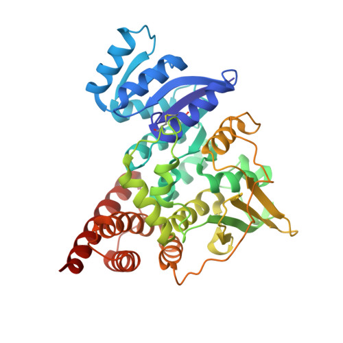
Crystal structure of 1-deoxy-D-xylulose-5-phosphate reductoisomerase, a crucial enzyme in the non-mevalonate pathway of isoprenoid biosynthesis.

Reuter, K.Sanderbrand, S.Jomaa, H.Wiesner, J.Steinbrecher, I.Beck, E.Hintz, M.Klebe, G.Stubbs, M.T.

(2002) J Biological Chem 277: 5378-5384

  • DOI: https://doi.org/10.1074/jbc.M109500200
  • Primary Citation Related Structures: 
    1K5H

  • PubMed Abstract: 

    We have solved the 2.5-A crystal structure of 1-deoxy-D-xylulose-5-phosphate reductoisomerase, an enzyme involved in the mevalonate-independent 2-C-methyl-D-erythritol-4-phosphate pathway of isoprenoid biosynthesis. The structure reveals that the enzyme is present as a homodimer. Each monomer displays a V-like shape and is composed of an amino-terminal dinucleotide binding domain, a connective domain, and a carboxyl-terminal four-helix bundle domain. The connective domain is responsible for dimerization and harbors most of the active site. The strictly conserved acidic residues Asp(150), Glu(152), Glu(231), and Glu(234) are clustered at the putative active site and are probably involved in the binding of divalent cations mandatory for enzyme activity. The connective and four-helix bundle domains show significant mobility upon superposition of the dinucleotide binding domains of the three conformational states present in the asymmetric unit of the crystal. A still more pronounced flexibility is observed for a loop spanning residues 186 to 216, which adopts two completely different conformations within the three protein conformers. A possible involvement of this loop in an induced fit during substrate binding is discussed.


  • Organizational Affiliation
    • Institut für Pharmazeutische Chemie, Philipps-Universität, Marbacher Weg 6, D-35032 Marburg, Germany. reuterk@mailer.uni-marburg.de

Macromolecule Content 

  • Total Structure Weight: 130.3 kDa 
  • Atom Count: 9,126 
  • Modeled Residue Count: 1,187 
  • Deposited Residue Count: 1,194 
  • Unique protein chains: 1

Macromolecules

Find similar proteins by:|  3D Structure
Entity ID: 1
MoleculeChains  Sequence LengthOrganismDetailsImage
1-deoxy-D-xylulose-5-phosphate reductoisomerase
A, B, C
398Escherichia coli K-12Mutation(s): 0 
Gene Names: dxr
EC: 1.1.1 (PDB Primary Data), 1.1.1.267 (UniProt)
UniProt
Find proteins for P45568 (Escherichia coli (strain K12))
Explore P45568 
Go to UniProtKB:  P45568
Entity Groups
Sequence Clusters30% Identity50% Identity70% Identity90% Identity95% Identity100% Identity
UniProt GroupP45568
Sequence Annotations
Expand
Reference Sequence

Experimental Data & Validation

Experimental Data

  • Method: X-RAY DIFFRACTION
  • Resolution: 2.50 Å
  • R-Value Free:  0.284 (Depositor), 0.283 (DCC) 
  • R-Value Work:  0.232 (Depositor), 0.233 (DCC) 
  • R-Value Observed: 0.232 (Depositor) 
Space Group: C 2 2 21
Unit Cell:
Length ( Å )Angle ( ˚ )
a = 101.266α = 90
b = 249.263β = 90
c = 132.112γ = 90
Software Package:
Software NamePurpose
DENZOdata reduction
SCALEPACKdata scaling
SOLVEphasing
CNSrefinement

Structure Validation

View Full Validation Report


Entry History 

Deposition Data

Revision History  (Full details and data files)

  • Version 1.0: 2002-02-27
    Type: Initial release
  • Version 1.1: 2008-04-27
    Changes: Version format compliance
  • Version 1.2: 2011-07-13
    Changes: Source and taxonomy, Version format compliance
  • Version 1.3: 2024-10-30
    Changes: Data collection, Database references, Structure summary