1K35 | pdb_00001k35

Crystal Structure of Phosphomannomutase/Phosphoglucomutase from P.aeruginosa


Experimental Data Snapshot

  • Method: X-RAY DIFFRACTION
  • Resolution: 2.20 Å
  • R-Value Free: 
    0.282 (Depositor) 
  • R-Value Work: 
    0.235 (Depositor) 
  • R-Value Observed: 
    0.238 (Depositor) 

wwPDB Validation 3D Report Full Report

Validation slider image for 1K35

This is version 1.3 of the entry. See complete history

Literature

Crystal structure of PMM/PGM: an enzyme in the biosynthetic pathway of P. aeruginosa virulence factors.

Regni, C.Tipton, P.A.Beamer, L.J.

(2002) Structure 10: 269-279

  • DOI: https://doi.org/10.1016/s0969-2126(02)00705-0
  • Primary Citation Related Structures: 
    1K2Y, 1K35

  • PubMed Abstract: 

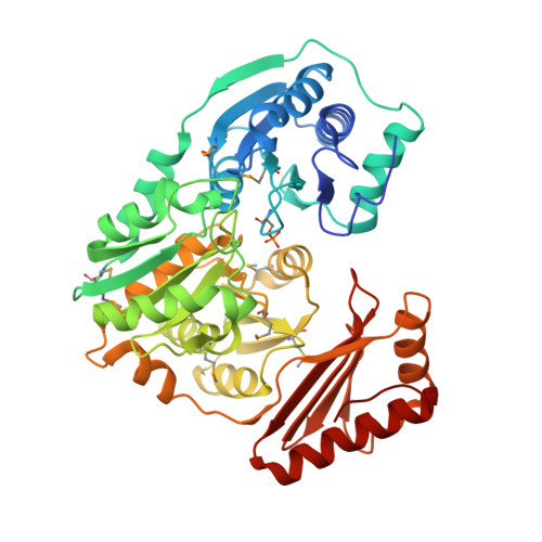
    The enzyme phosphomannomutase/phosphoglucomutase (PMM/PGM) from P. aeruginosa is required for the biosynthesis of two bacterial exopolysaccharides: alginate and lipopolysaccharide (LPS). Both of these molecules play a role in the virulence of P. aeruginosa, an important human pathogen known for its ability to develop antibiotic resistance and cause chronic lung infections in cystic fibrosis patients. The crystal structure of PMM/PGM shows that the enzyme has four domains, three of which have a similar three-dimensional fold. Residues from all four domains of the protein contribute to the formation of a large active site cleft in the center of the molecule. Detailed information on the active site of PMM/PGM lays the foundation for structure-based inhibitor design. Inhibitors of sufficient potency and specificity should impair the biosynthesis of alginate and LPS, and may facilitate clearance of the bacteria by the host immune system and increase the efficacy of conventional antibiotic treatment against chronic P. aeruginosa infections.


  • Organizational Affiliation
    • Department of Biochemistry, 117 Schweitzer Hall, University of Missouri-Columbia, Columbia, MO 65211, USA.

Macromolecule Content 

  • Total Structure Weight: 50.87 kDa 
  • Atom Count: 3,547 
  • Modeled Residue Count: 455 
  • Deposited Residue Count: 463 
  • Unique protein chains: 1

Macromolecules

Find similar proteins by:|  3D Structure
Entity ID: 1
MoleculeChains  Sequence LengthOrganismDetailsImage
Phosphomannomutase463Pseudomonas aeruginosaMutation(s): 9 
Gene Names: AlgC
EC: 5.4.2.8 (PDB Primary Data), 5.4.2.2 (UniProt)
UniProt
Find proteins for P26276 (Pseudomonas aeruginosa (strain ATCC 15692 / DSM 22644 / CIP 104116 / JCM 14847 / LMG 12228 / 1C / PRS 101 / PAO1))
Explore P26276 
Go to UniProtKB:  P26276
Entity Groups
Sequence Clusters30% Identity50% Identity70% Identity90% Identity95% Identity100% Identity
UniProt GroupP26276
Sequence Annotations
Expand
Reference Sequence

Small Molecules

Ligands 1 Unique
IDChains Name / Formula / InChI Key2D Diagram3D Interactions
ZN

Query on ZN


Download:Ideal Coordinates CCD File
B [auth A]ZINC ION
Zn
PTFCDOFLOPIGGS-UHFFFAOYSA-N
Modified Residues  2 Unique
IDChains TypeFormula2D DiagramParent
MSE
Query on MSE
A
L-PEPTIDE LINKINGC5 H11 N O2 SeMET
SEP
Query on SEP
A
L-PEPTIDE LINKINGC3 H8 N O6 PSER

Experimental Data & Validation

Experimental Data

  • Method: X-RAY DIFFRACTION
  • Resolution: 2.20 Å
  • R-Value Free:  0.282 (Depositor) 
  • R-Value Work:  0.235 (Depositor) 
  • R-Value Observed: 0.238 (Depositor) 
Space Group: P 21 21 21
Unit Cell:
Length ( Å )Angle ( ˚ )
a = 71.038α = 90
b = 73.263β = 90
c = 92.354γ = 90
Software Package:
Software NamePurpose
SOLVEphasing
REFMACrefinement
DENZOdata reduction
SCALEPACKdata scaling

Structure Validation

View Full Validation Report


Entry History 

Deposition Data

Revision History  (Full details and data files)

  • Version 1.0: 2002-02-13
    Type: Initial release
  • Version 1.1: 2008-04-27
    Changes: Version format compliance
  • Version 1.2: 2011-07-13
    Changes: Version format compliance
  • Version 1.3: 2024-11-20
    Changes: Data collection, Database references, Derived calculations, Structure summary