1K2D | pdb_00001k2d

Crystal structure of the autoimmune MHC class II I-Au complexed with myelin basic protein 1-11 at 2.2A


Experimental Data Snapshot

  • Method: X-RAY DIFFRACTION
  • Resolution: 2.20 Å
  • R-Value Free: 
    0.276 (Depositor) 
  • R-Value Work: 
    0.248 (Depositor) 
  • R-Value Observed: 
    0.249 (Depositor) 

Starting Model: other
View more details

wwPDB Validation 3D Report Full Report

Validation slider image for 1K2D

This is version 2.3 of the entry. See complete history

Literature

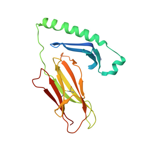
Structural snapshot of aberrant antigen presentation linked to autoimmunity: the immunodominant epitope of MBP complexed with I-Au

He, X.L.Radu, C.Sidney, J.Sette, A.Ward, E.S.Garcia, K.C.

(2002) Immunity 17: 83-94

  • DOI: https://doi.org/10.1016/s1074-7613(02)00340-0
  • Primary Citation Related Structures: 
    1K2D

  • PubMed Abstract: 

    Murine experimental allergic encephalomyelitis (EAE) is a useful model for the demyelinating, autoimmune disease multiple sclerosis. In the EAE system, the immunodominant N-terminal epitope of myelin basic protein (MBP) is an unusually short, weakly binding peptide antigen which elicits highly biased TCR chain usage. In the 2.2 A crystal structure of I-A(u)/MBP1-11 complex, only MBP residues 1-7 are bound toward one end of the peptide binding cleft. The fourth residue of MBP1-11 is located in an incompatible p6 pocket of I-A(u), thus explaining the short half-life of I-A(u) complexed with Ac1-11. MBP peptides extended at the C terminus of Ac1-11 result in dramatic affinity increases, likely attributed to register shifting to a higher affinity cryptic epitope, which could potentially mask the presentation of the immunodominant MBP1-11 peptide during thymic education.


  • Organizational Affiliation
    • Department of Microbiology and Immunology, Stanford University School of Medicine, CA 94305, USA.

Macromolecule Content 

  • Total Structure Weight: 46.95 kDa 
  • Atom Count: 3,597 
  • Modeled Residue Count: 379 
  • Deposited Residue Count: 401 
  • Unique protein chains: 3

Macromolecules

Find similar proteins by:|  3D Structure
Entity ID: 1
MoleculeChains  Sequence LengthOrganismDetailsImage
H-2 class II histocompatibility antigen, A-U alpha chain189Mus musculusMutation(s): 0 
UniProt
Find proteins for P14438 (Mus musculus)
Explore P14438 
Go to UniProtKB:  P14438
Entity Groups
Sequence Clusters30% Identity50% Identity70% Identity90% Identity95% Identity100% Identity
UniProt GroupP14438
Glycosylation
Glycosylation Sites: 2
Sequence Annotations
Expand
Reference Sequence
Find similar proteins by:|  3D Structure
Entity ID: 2
MoleculeChains  Sequence LengthOrganismDetailsImage
H-2 class II histocompatibility antigen, A-U beta chain189Mus musculusMutation(s): 0 
UniProt
Find proteins for P06344 (Mus musculus)
Explore P06344 
Go to UniProtKB:  P06344
Entity Groups
Sequence Clusters30% Identity50% Identity70% Identity90% Identity95% Identity100% Identity
UniProt GroupP06344
Glycosylation
Glycosylation Sites: 1Go to GlyGen: P06344-1
Sequence Annotations
Expand
Reference Sequence
Find similar proteins by:  Sequence   |   3D Structure  
Entity ID: 3
MoleculeChains  Sequence LengthOrganismDetailsImage
Myelin Basic Protein peptide with 8 residue linker peptideC [auth P]23N/AMutation(s): 0 
UniProt & NIH Common Fund Data Resources
Find proteins for P02686 (Homo sapiens)
Explore P02686 
Go to UniProtKB:  P02686
PHAROS:  P02686
GTEx:  ENSG00000197971 
Entity Groups
Sequence Clusters30% Identity50% Identity70% Identity90% Identity95% Identity100% Identity
UniProt GroupP02686
Sequence Annotations
Expand
Reference Sequence

Experimental Data & Validation

Experimental Data

  • Method: X-RAY DIFFRACTION
  • Resolution: 2.20 Å
  • R-Value Free:  0.276 (Depositor) 
  • R-Value Work:  0.248 (Depositor) 
  • R-Value Observed: 0.249 (Depositor) 
Space Group: C 2 2 21
Unit Cell:
Length ( Å )Angle ( ˚ )
a = 99.15α = 90
b = 110.59β = 90
c = 94.84γ = 90
Software Package:
Software NamePurpose
MOSFLMdata reduction
SCALAdata scaling
AMoREphasing
CNSrefinement
CCP4data scaling

Structure Validation

View Full Validation Report


Entry History 

Deposition Data

Revision History  (Full details and data files)

  • Version 1.0: 2003-03-04
    Type: Initial release
  • Version 1.1: 2008-04-27
    Changes: Version format compliance
  • Version 1.2: 2011-07-13
    Changes: Non-polymer description, Version format compliance
  • Version 2.0: 2020-07-29
    Type: Remediation
    Reason: Carbohydrate remediation
    Changes: Advisory, Atomic model, Data collection, Database references, Derived calculations, Non-polymer description, Structure summary
  • Version 2.1: 2022-12-21
    Changes: Database references, Structure summary
  • Version 2.2: 2024-04-03
    Changes: Data collection, Refinement description
  • Version 2.3: 2024-10-16
    Changes: Structure summary