1JXA | pdb_00001jxa

GLUCOSAMINE 6-PHOSPHATE SYNTHASE WITH GLUCOSE 6-PHOSPHATE


Experimental Data Snapshot

  • Method: X-RAY DIFFRACTION
  • Resolution: 3.10 Å
  • R-Value Free: 
    0.280 (Depositor) 
  • R-Value Work: 
    0.200 (Depositor), 0.213 (DCC) 
  • R-Value Observed: 
    0.220 (Depositor) 

Starting Models: experimental
View more details

wwPDB Validation 3D Report Full Report

Validation slider image for 1JXA

Ligand Structure Quality Assessment 


This is version 1.5 of the entry. See complete history

Literature

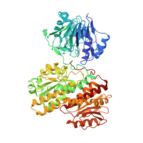
Channeling of ammonia in glucosamine-6-phosphate synthase.

Teplyakov, A.Obmolova, G.Badet, B.Badet-Denisot, M.A.

(2001) J Mol Biology 313: 1093-1102

  • DOI: https://doi.org/10.1006/jmbi.2001.5094
  • Primary Citation Related Structures: 
    1JXA

  • PubMed Abstract: 

    Glucosamine-6-phosphate synthase catalyses the first and rate-limiting step in hexosamine metabolism, converting fructose 6-phosphate into glucosamine 6-phosphate in the presence of glutamine. The crystal structure of the Escherichia coli enzyme reveals the domain organisation of the homodimeric molecule. The 18 A hydrophobic channel sequestered from the solvent connects the glutaminase and isomerase active sites, and provides a means of ammonia transfer from glutamine to sugar phosphate. The C-terminal decapeptide sandwiched between the two domains plays a central role in the transfer. Based on the structure, a mechanism of enzyme action and self-regulation is proposed. It involves large domain movements triggered by substrate binding that lead to the formation of the channel.


  • Organizational Affiliation
    • European Molecular Biology Laboratory, Notkestr. 85, D-22603 Hamburg, Germany. alexey@carb.inst.gov

Macromolecule Content 

  • Total Structure Weight: 200.97 kDa 
  • Atom Count: 14,156 
  • Modeled Residue Count: 1,824 
  • Deposited Residue Count: 1,824 
  • Unique protein chains: 1

Macromolecules

Find similar proteins by:|  3D Structure
Entity ID: 1
MoleculeChains  Sequence LengthOrganismDetailsImage
glucosamine 6-phosphate synthase
A, B, C
608Escherichia coliMutation(s): 0 
EC: 2.6.1.16
UniProt
Find proteins for P17169 (Escherichia coli (strain K12))
Explore P17169 
Go to UniProtKB:  P17169
Entity Groups
Sequence Clusters30% Identity50% Identity70% Identity90% Identity95% Identity100% Identity
UniProt GroupP17169
Sequence Annotations
Expand
Reference Sequence

Experimental Data & Validation

Experimental Data

  • Method: X-RAY DIFFRACTION
  • Resolution: 3.10 Å
  • R-Value Free:  0.280 (Depositor) 
  • R-Value Work:  0.200 (Depositor), 0.213 (DCC) 
  • R-Value Observed: 0.220 (Depositor) 
Space Group: C 1 2 1
Unit Cell:
Length ( Å )Angle ( ˚ )
a = 131.4α = 90
b = 112.4β = 96.4
c = 185.1γ = 90
Software Package:
Software NamePurpose
MOLREPphasing
REFMACrefinement
DENZOdata reduction
SCALEPACKdata scaling

Structure Validation

View Full Validation Report


Ligand Structure Quality Assessment 


Entry History 

Deposition Data

Revision History  (Full details and data files)

  • Version 1.0: 2001-11-21
    Type: Initial release
  • Version 1.1: 2008-04-27
    Changes: Version format compliance
  • Version 1.2: 2011-07-13
    Changes: Version format compliance
  • Version 1.3: 2018-10-03
    Changes: Data collection
  • Version 1.4: 2023-08-16
    Changes: Data collection, Database references, Derived calculations, Refinement description
  • Version 1.5: 2024-12-25
    Changes: Advisory, Derived calculations, Structure summary