1JVV | pdb_00001jvv

CRYSTAL STRUCTURE OF RIBONUCLEASE A (RETRO-SOAKED FORM)


Experimental Data Snapshot

  • Method: X-RAY DIFFRACTION
  • Resolution: 2.20 Å
  • R-Value Free: 
    0.260 (Depositor) 
  • R-Value Work: 
    0.170 (Depositor) 
  • R-Value Observed: 
    0.180 (Depositor) 

Starting Model: experimental
View more details

wwPDB Validation 3D Report Full Report

Validation slider image for 1JVV

This is version 1.4 of the entry. See complete history

Literature

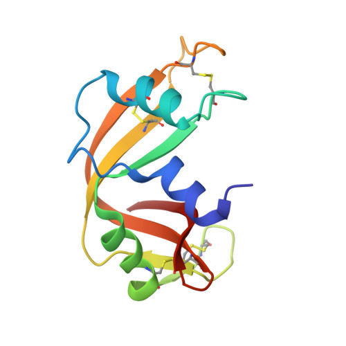
Reversible Substrate-Induced Domain Motions in Ribonuclease A

Vitagliano, L.Merlino, A.Zagari, A.Mazzarella, L.

(2002) Proteins 46: 97-104

  • DOI: https://doi.org/10.1002/prot.10033
  • Primary Citation Related Structures: 
    1JVT, 1JVU, 1JVV

  • PubMed Abstract: 

    Despite the increasing number of successful determinations of complex protein structures the understanding of their dynamics properties is still rather limited. Using X-ray crystallography, we demonstrate that ribonuclease A (RNase A) undergoes significant domain motions upon ligand binding. In particular, when cytidine 2'-monophosphate binds to RNase A, the structure of the enzyme becomes more compact. Interestingly, our data also show that these structural alterations are fully reversible in the crystal state. These findings provide structural bases for the dynamic behavior of RNase A in the binding of the substrate shown by Petsko and coworkers (Rasmussen et al. Nature 1992;357:423-424). These subtle domain motions may assume functional relevance for more complex system and may play a significant role in the cooperativity of oligomeric enzymes.


  • Organizational Affiliation
    • Centro di Biocristallografia, CNR, Napoli, Italy.

Macromolecule Content 

  • Total Structure Weight: 27.42 kDa 
  • Atom Count: 1,963 
  • Modeled Residue Count: 248 
  • Deposited Residue Count: 248 
  • Unique protein chains: 1

Macromolecules

Find similar proteins by:|  3D Structure
Entity ID: 1
MoleculeChains  Sequence LengthOrganismDetailsImage
RIBONUCLEASE A
A, B
124Bos taurusMutation(s): 0 
EC: 3.1.27.5 (PDB Primary Data), 4.6.1.18 (UniProt)
UniProt
Find proteins for P61823 (Bos taurus)
Explore P61823 
Go to UniProtKB:  P61823
Entity Groups
Sequence Clusters30% Identity50% Identity70% Identity90% Identity95% Identity100% Identity
UniProt GroupP61823
Sequence Annotations
Expand
Reference Sequence

Experimental Data & Validation

Experimental Data

  • Method: X-RAY DIFFRACTION
  • Resolution: 2.20 Å
  • R-Value Free:  0.260 (Depositor) 
  • R-Value Work:  0.170 (Depositor) 
  • R-Value Observed: 0.180 (Depositor) 
Space Group: C 1 2 1
Unit Cell:
Length ( Å )Angle ( ˚ )
a = 101.27α = 90
b = 33.2β = 90.1
c = 73.58γ = 90
Software Package:
Software NamePurpose
DENZOdata reduction
SCALEPACKdata scaling
X-PLORmodel building
X-PLORrefinement
X-PLORphasing

Structure Validation

View Full Validation Report


Entry History 

Deposition Data

Revision History  (Full details and data files)

  • Version 1.0: 2002-06-05
    Type: Initial release
  • Version 1.1: 2008-04-27
    Changes: Version format compliance
  • Version 1.2: 2011-07-13
    Changes: Version format compliance
  • Version 1.3: 2023-08-16
    Changes: Data collection, Database references, Refinement description
  • Version 1.4: 2024-10-16
    Changes: Structure summary