1JV5 | pdb_00001jv5

Anti-blood group A Fv


Experimental Data Snapshot

  • Method: X-RAY DIFFRACTION
  • Resolution: 2.20 Å
  • R-Value Free: 
    0.223 (Depositor), 0.212 (DCC) 
  • R-Value Work: 
    0.183 (Depositor), 0.209 (DCC) 
  • R-Value Observed: 
    0.183 (Depositor) 

wwPDB Validation 3D Report Full Report

Validation slider image for 1JV5

This is version 1.5 of the entry. See complete history

Literature

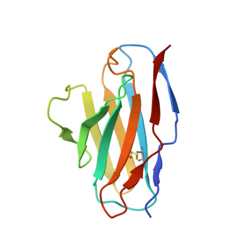
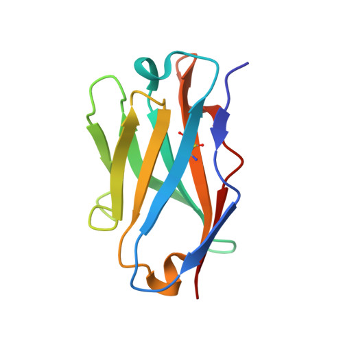
Structure of an anti-blood group A Fv and improvement of its binding affinity without loss of specificity.

Thomas, R.Patenaude, S.I.MacKenzie, C.R.To, R.Hirama, T.Young, N.M.Evans, S.V.

(2002) J Biological Chem 277: 2059-2064

  • DOI: https://doi.org/10.1074/jbc.M104364200
  • Primary Citation Related Structures: 
    1JV5

  • PubMed Abstract: 

    The specificity of antibody recognition of the ABO blood group trisaccharide antigens has been explored by crystal structure analysis and mutation methods. The crystal structure of the Fv corresponding to the anti-blood group A antibody AC1001 has been determined to 2.2-A resolution and reveals a binding pocket that is complementary to the blood group A-trisaccharide antigen. The effect of mutating specific residues lining this pocket on binding to the A and B blood group oligosaccharide antigens was investigated through a panel of single point mutations and through a phage library of mutations in complementarity determining region H3. Both approaches gave several mutants with improved affinity for antigen. Surface plasmon resonance indicated up to 8-fold enhancement in affinity for the A-pentasaccharide with no observable binding to the blood group B antigen. This is the first example of single point mutations in a carbohydrate-binding antibody resulting in significant increases in binding affinity without loss of specificity.


  • Organizational Affiliation
    • Department of Biochemistry, Microbiology and Immunology, University of Ottawa, Ottawa, Ontario K1H 8M5, Canada.

Macromolecule Content 

  • Total Structure Weight: 24.69 kDa 
  • Atom Count: 1,802 
  • Modeled Residue Count: 224 
  • Deposited Residue Count: 224 
  • Unique protein chains: 2

Macromolecules

Find similar proteins by:|  3D Structure
Entity ID: 1
MoleculeChains  Sequence LengthOrganismDetailsImage
Ig kappa chain precursor V region107Homo sapiensMutation(s): 0 
UniProt & NIH Common Fund Data Resources
Find proteins for P01594 (Homo sapiens)
Explore P01594 
Go to UniProtKB:  P01594
GTEx:  ENSG00000242076 
Entity Groups
Sequence Clusters30% Identity50% Identity70% Identity90% Identity95% Identity100% Identity
UniProt GroupP01594
Sequence Annotations
Expand
Reference Sequence
Find similar proteins by:|  3D Structure
Entity ID: 2
MoleculeChains  Sequence LengthOrganismDetailsImage
Ig chain heavy chain precursor V region117Homo sapiensMutation(s): 0 

Experimental Data & Validation

Experimental Data

  • Method: X-RAY DIFFRACTION
  • Resolution: 2.20 Å
  • R-Value Free:  0.223 (Depositor), 0.212 (DCC) 
  • R-Value Work:  0.183 (Depositor), 0.209 (DCC) 
  • R-Value Observed: 0.183 (Depositor) 
Space Group: P 21 21 21
Unit Cell:
Length ( Å )Angle ( ˚ )
a = 51.71α = 90
b = 57.96β = 90
c = 83.58γ = 90
Software Package:
Software NamePurpose
CAD4data collection
ROTAVATAdata reduction
MERLOTphasing
X-PLORrefinement
CAD4data reduction
CCP4data scaling
ROTAVATAdata scaling

Structure Validation

View Full Validation Report


Entry History 

Deposition Data

Revision History  (Full details and data files)

  • Version 1.0: 2002-01-09
    Type: Initial release
  • Version 1.1: 2008-04-27
    Changes: Version format compliance
  • Version 1.2: 2011-07-13
    Changes: Version format compliance
  • Version 1.3: 2017-10-04
    Changes: Refinement description
  • Version 1.4: 2018-04-18
    Changes: Data collection
  • Version 1.5: 2024-10-09
    Changes: Data collection, Database references, Structure summary