1JQ7 | pdb_00001jq7

HCMV protease dimer-interface mutant, S225Y complexed to Inhibitor BILC 408


Experimental Data Snapshot

  • Method: X-RAY DIFFRACTION
  • Resolution: 3.00 Å
  • R-Value Free: 
    0.339 (Depositor) 
  • R-Value Work: 
    0.260 (Depositor) 

Starting Model: experimental
View more details

wwPDB Validation 3D Report Full Report

Validation slider image for 1JQ7

This is version 1.7 of the entry. See complete history

Literature

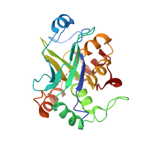
Molecular mechanism for dimerization to regulate the catalytic activity of human cytomegalovirus protease.

Batra, R.Khayat, R.Tong, L.

(2001) Nat Struct Biol 8: 810-817

  • DOI: https://doi.org/10.1038/nsb0901-810
  • Primary Citation Related Structures: 
    1JQ6, 1JQ7

  • PubMed Abstract: 

    Biochemical studies indicate that dimerization is required for the catalytic activity of herpesvirus proteases, whereas structural studies show a complete active site in each monomer, away from the dimer interface. Here we report kinetic, biophysical and crystallographic characterizations of structure-based mutants in the dimer interface of human cytomegalovirus (HCMV) protease. Such mutations can produce a 1,700-fold reduction in the kcat while having minimal effects on the K(m). Dimer stability is not affected by these mutations, suggesting that dimerization itself is insufficient for activity. There are large changes in monomer conformation and dimer organization of the apo S225Y mutant enzyme. However, binding of an activated peptidomimetic inhibitor induced a conformation remarkably similar to the wild type protease. Our studies suggest that appropriate dimer formation may be required to indirectly stabilize the protease oxyanion hole, revealing a novel mechanism for dimerization to regulate enzyme activity.


  • Organizational Affiliation
    • Department of Biological Sciences, Columbia University, New York, New York 10027, USA.

Macromolecule Content 

  • Total Structure Weight: 57.85 kDa 
  • Atom Count: 3,395 
  • Modeled Residue Count: 427 
  • Deposited Residue Count: 512 
  • Unique protein chains: 1

Macromolecules

Find similar proteins by:|  3D Structure
Entity ID: 1
MoleculeChains  Sequence LengthOrganismDetailsImage
ASSEMBLIN
A, B
256Human betaherpesvirus 5Mutation(s): 2 
EC: 3.4.21.97
UniProt
Find proteins for P16753 (Human cytomegalovirus (strain AD169))
Explore P16753 
Go to UniProtKB:  P16753
Entity Groups
Sequence Clusters30% Identity50% Identity70% Identity90% Identity95% Identity100% Identity
UniProt GroupP16753
Sequence Annotations
Expand
Reference Sequence

Small Molecules

Ligands 1 Unique
IDChains Name / Formula / InChI Key2D Diagram3D Interactions
0FP

Query on 0FP


Download:Ideal Coordinates CCD File
C [auth A],
D [auth B]
N-(6-aminohexanoyl)-3-methyl-L-valyl-3-methyl-L-valyl-N~1~-[(2S,3S)-3-hydroxy-4-oxo-4-{[(1R)-1-phenylpropyl]amino}butan-2-yl]-N~4~,N~4~-dimethyl-L-aspartamide
C37 H63 N7 O7
RICYNZHPHSJRNO-GYZAPQSVSA-N

Experimental Data & Validation

Experimental Data

  • Method: X-RAY DIFFRACTION
  • Resolution: 3.00 Å
  • R-Value Free:  0.339 (Depositor) 
  • R-Value Work:  0.260 (Depositor) 
Space Group: P 41 21 2
Unit Cell:
Length ( Å )Angle ( ˚ )
a = 74.25α = 90
b = 74.25β = 90
c = 215.8γ = 90
Software Package:
Software NamePurpose
COMOphasing
CNSrefinement
DENZOdata reduction
SCALEPACKdata scaling

Structure Validation

View Full Validation Report


Entry History 

Deposition Data

Revision History  (Full details and data files)

  • Version 1.0: 2001-09-12
    Type: Initial release
  • Version 1.1: 2008-04-27
    Changes: Version format compliance
  • Version 1.2: 2011-07-13
    Changes: Atomic model, Database references, Derived calculations, Non-polymer description, Structure summary, Version format compliance
  • Version 1.3: 2012-12-12
    Changes: Other
  • Version 1.4: 2021-10-27
    Changes: Database references, Derived calculations
  • Version 1.5: 2023-08-16
    Changes: Data collection, Refinement description
  • Version 1.6: 2024-03-13
    Changes: Source and taxonomy, Structure summary
  • Version 1.7: 2024-11-20
    Changes: Structure summary