1JMX | pdb_00001jmx

crystal structure of a quinohemoprotein amine dehydrogenase from pseudomonas putida


Experimental Data Snapshot

  • Method: X-RAY DIFFRACTION
  • Resolution: 1.90 Å
  • R-Value Free: 
    0.267 (Depositor) 
  • R-Value Work: 
    0.209 (Depositor) 

wwPDB Validation 3D Report Full Report

Validation slider image for 1JMX

This is version 1.3 of the entry. See complete history

Literature

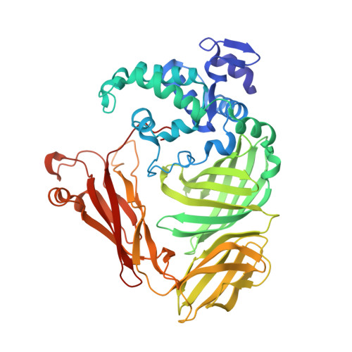
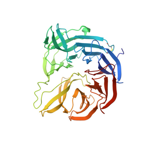
Crystal structure of quinohemoprotein amine dehydrogenase from Pseudomonas putida. Identification of a novel quinone cofactor encaged by multiple thioether cross-bridges.

Satoh, A.Kim, J.K.Miyahara, I.Devreese, B.Vandenberghe, I.Hacisalihoglu, A.Okajima, T.Kuroda, S.Adachi, O.Duine, J.A.Van Beeumen, J.Tanizawa, K.Hirotsu, K.

(2002) J Biological Chem 277: 2830-2834

  • DOI: https://doi.org/10.1074/jbc.M109090200
  • Primary Citation Related Structures: 
    1JMX, 1JMZ

  • PubMed Abstract: 

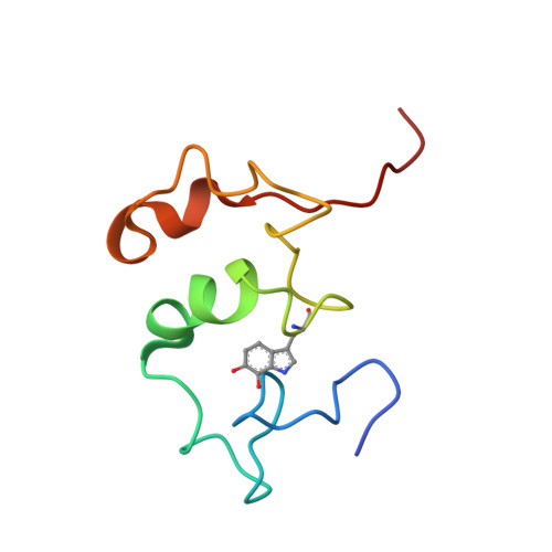
    The crystal structure of a quinohemoprotein amine dehydrogenase from Pseudomonas putida has been determined at 1.9-A resolution. The enzyme comprises three non-identical subunits: a four-domain alpha-subunit that harbors a di-heme cytochrome c, a seven-bladed beta-propeller beta-subunit that provides part of the active site, and a small gamma-subunit that contains a novel cross-linked, proteinous quinone cofactor, cysteine tryptophylquinone. More surprisingly, the catalytic gamma-subunit contains three additional chemical cross-links that encage the cysteine tryptophylquinone cofactor, involving a cysteine side chain bridged to either an Asp or Glu residue all in a hitherto unknown thioether bonding with a methylene carbon atom of acidic amino acid side chains. Thus, the structure of the 79-residue gamma-subunit is quite unusual, containing four internal cross-links in such a short polypeptide chain that would otherwise be difficult to fold into a globular structure.


  • Organizational Affiliation
    • Department of Chemistry, Graduate School of Science, Osaka City University, Osaka 558-8585, Japan.

Macromolecule Content 

  • Total Structure Weight: 103.2 kDa 
  • Atom Count: 7,611 
  • Modeled Residue Count: 909 
  • Deposited Residue Count: 922 
  • Unique protein chains: 3

Macromolecules

Find similar proteins by:|  3D Structure
Entity ID: 1
MoleculeChains  Sequence LengthOrganismDetailsImage
Amine Dehydrogenase494Pseudomonas putidaMutation(s): 0 
UniProt
Find proteins for Q8VW85 (Pseudomonas putida)
Explore Q8VW85 
Go to UniProtKB:  Q8VW85
Entity Groups
Sequence Clusters30% Identity50% Identity70% Identity90% Identity95% Identity100% Identity
UniProt GroupQ8VW85
Sequence Annotations
Expand
Reference Sequence
Find similar proteins by:|  3D Structure
Entity ID: 2
MoleculeChains  Sequence LengthOrganismDetailsImage
Amine Dehydrogenase349Pseudomonas putidaMutation(s): 0 
UniProt
Find proteins for Q8VW82 (Pseudomonas putida)
Explore Q8VW82 
Go to UniProtKB:  Q8VW82
Entity Groups
Sequence Clusters30% Identity50% Identity70% Identity90% Identity95% Identity100% Identity
UniProt GroupQ8VW82
Sequence Annotations
Expand
Reference Sequence
Find similar proteins by:|  3D Structure
Entity ID: 3
MoleculeChains  Sequence LengthOrganismDetailsImage
Amine DehydrogenaseC [auth G]79Pseudomonas putidaMutation(s): 0 
EC: 1.4.9
UniProt
Find proteins for P0A182 (Pseudomonas putida)
Explore P0A182 
Go to UniProtKB:  P0A182
Entity Groups
Sequence Clusters30% Identity50% Identity70% Identity90% Identity95% Identity100% Identity
UniProt GroupP0A182
Sequence Annotations
Expand
Reference Sequence

Small Molecules

Modified Residues  1 Unique
IDChains TypeFormula2D DiagramParent
TRQ
Query on TRQ
C [auth G]L-PEPTIDE LINKINGC11 H10 N2 O4TRP

Experimental Data & Validation

Experimental Data

  • Method: X-RAY DIFFRACTION
  • Resolution: 1.90 Å
  • R-Value Free:  0.267 (Depositor) 
  • R-Value Work:  0.209 (Depositor) 
Space Group: C 1 2 1
Unit Cell:
Length ( Å )Angle ( ˚ )
a = 167.21α = 90
b = 92.37β = 112
c = 79.3γ = 90
Software Package:
Software NamePurpose
MOSFLMdata reduction
SCALAdata scaling
SHARPphasing
X-PLORrefinement
CCP4data scaling

Structure Validation

View Full Validation Report


Entry History 

Deposition Data

Revision History  (Full details and data files)

  • Version 1.0: 2002-01-16
    Type: Initial release
  • Version 1.1: 2008-04-27
    Changes: Version format compliance
  • Version 1.2: 2011-07-13
    Changes: Version format compliance
  • Version 1.3: 2025-03-26
    Changes: Data collection, Database references, Derived calculations, Structure summary