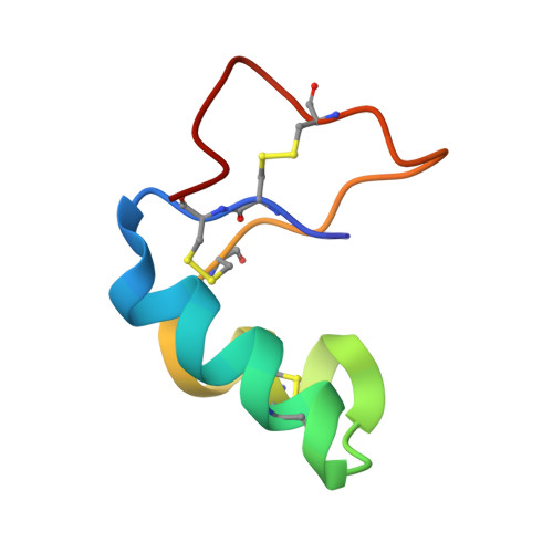
Comparative membrane interaction study of viscotoxins A3, A2 and B from mistletoe (Viscum album) and connections with their structures
Coulon, A., Mosbah, A., Lopez, A., Sautereau, A.M., Schaller, G., Urech, K., Rouge, P., Darbon, H.(2003) Biochem J 374: 71-78
- PubMed: 12733989
- DOI: https://doi.org/10.1042/BJ20030488
- Primary Citation of Related Structures:
1JMP - PubMed Abstract:
Viscotoxins A2 (VA2) and B (VB) are, together with viscotoxin A3 (VA3), among the most abundant viscotoxin isoforms that occur in mistletoe-derived medicines used in anti-cancer therapy. Although these isoforms have a high degree of amino-acid-sequence similarity, they are strikingly different from each other in their in vitro cytotoxic potency towards tumour cells. First, as VA3 is the only viscotoxin whose three-dimensional (3D) structure has been solved to date, we report the NMR determination of the 3D structures of VA2 and VB. Secondly, to account for the in vitro cytotoxicity discrepancy, we carried out a comparative study of the interaction of the three viscotoxins with model membranes. Although the overall 3D structure is highly conserved among the three isoforms, some discrete structural features and associated surface properties readily account for the different affinity and perturbation of model membranes. VA3 and VA2 interact in a similar way, but the weaker hydrophobic character of VA2 is thought to be mainly responsible for the apparent different affinity towards membranes. VB is much less active than the other two viscotoxins and does not insert into model membranes. This could be related to the occurrence of a single residue (Arg25) protruding outside the hydrophobic plane formed by the two amphipathic alpha-helices, through which viscotoxins are supposed to interact with plasma membranes.
Organizational Affiliation:
Institut de Pharmacologie et de Biologie Structurale, Centre National de la Recherche Scientifique UMR 5089, 205 route de Narbonne, 31077 Toulouse 4, France.


