1JJH | pdb_00001jjh

E2 DNA-binding Domain from Bovine Papillomavirus Type 1


Experimental Data Snapshot

  • Method: X-RAY DIFFRACTION
  • Resolution: 2.50 Å
  • R-Value Free: 
    0.266 (Depositor) 
  • R-Value Work: 
    0.225 (Depositor) 
  • R-Value Observed: 
    0.225 (Depositor) 

wwPDB Validation 3D Report Full Report

Validation slider image for 1JJH

This is version 1.4 of the entry. See complete history

Literature

Subunit rearrangement accompanies sequence-specific DNA binding by the bovine papillomavirus-1 E2 protein.

Hegde, R.S.Wang, A.F.Kim, S.S.Schapira, M.

(1998) J Mol Biology 276: 797-808

  • DOI: https://doi.org/10.1006/jmbi.1997.1587
  • Primary Citation Related Structures: 
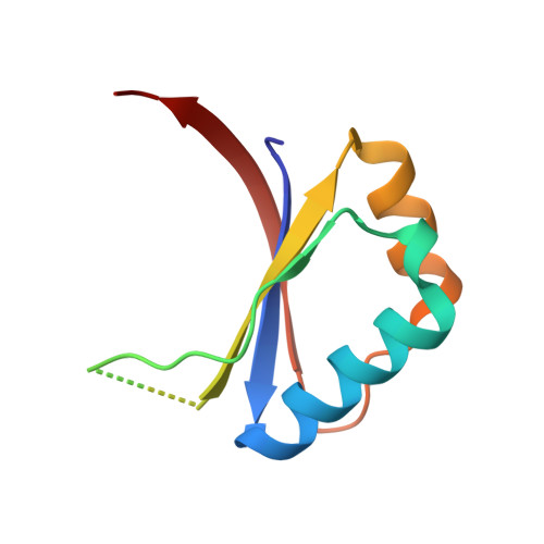
    1JJH

  • PubMed Abstract: 

    The 2.5 A crystal structures of the DNA-binding domain of the E2 protein from bovine papillomavirus strain 1 and its complex with DNA are presented. E2 is a transcriptional regulatory protein that is also involved in viral DNA replication. It is the structural prototype for a novel class of DNA-binding proteins: dimeric beta-barrels with surface alpha-helices that serve as recognition helices. These helices contain the amino-acid residues involved in sequence-specifying interactions. The E2 proteins from different papillomavirus strains recognize and bind to the same consensus 12 base-pair DNA sequence. However, recent evidence from solution studies points to differences in the mechanisms by which E2 from the related viral strains bovine papillomavirus-1 and human papillomavirus-16 discriminate between DNA targets based on non-contacted nucleotide sequences. This report provides evidence that sequence-specific DNA-binding is accompanied by a rearrangement of protein subunits and deformation of the DNA. These results suggest that, along with DNA sequence-dependent conformational properties, protein subunit orientation plays a significant role in the mechanisms of target selection utilized by E2.


  • Organizational Affiliation
    • Department of Biochemistry and Program in Structural Biology, Skirball Institute of Biomolecular Medicine, New York University Medical Center, 540 First Avenue, New York, NY 10016, USA.

Macromolecule Content 

  • Total Structure Weight: 29.08 kDa 
  • Atom Count: 1,896 
  • Modeled Residue Count: 228 
  • Deposited Residue Count: 258 
  • Unique protein chains: 1

Macromolecules

Find similar proteins by:|  3D Structure
Entity ID: 1
MoleculeChains  Sequence LengthOrganismDetailsImage
REGULATORY PROTEIN E2
A, B, C
86Deltapapillomavirus 4Mutation(s): 0 
Gene Names: E2
UniProt
Find proteins for P03122 (Bovine papillomavirus type 1)
Explore P03122 
Go to UniProtKB:  P03122
Entity Groups
Sequence Clusters30% Identity50% Identity70% Identity90% Identity95% Identity100% Identity
UniProt GroupP03122
Sequence Annotations
Expand
Reference Sequence

Experimental Data & Validation

Experimental Data

  • Method: X-RAY DIFFRACTION
  • Resolution: 2.50 Å
  • R-Value Free:  0.266 (Depositor) 
  • R-Value Work:  0.225 (Depositor) 
  • R-Value Observed: 0.225 (Depositor) 
Space Group: P 65 2 2
Unit Cell:
Length ( Å )Angle ( ˚ )
a = 122.29α = 90
b = 122.29β = 90
c = 85.019γ = 120
Software Package:
Software NamePurpose
DENZOdata reduction
SCALEPACKdata scaling
AMoREphasing
CNSrefinement

Structure Validation

View Full Validation Report


Entry History 

Deposition Data

Revision History  (Full details and data files)

  • Version 1.0: 2001-07-18
    Type: Initial release
  • Version 1.1: 2008-04-27
    Changes: Version format compliance
  • Version 1.2: 2011-07-13
    Changes: Version format compliance
  • Version 1.3: 2017-10-04
    Changes: Refinement description
  • Version 1.4: 2024-11-13
    Changes: Data collection, Database references, Structure summary