1JIN | pdb_00001jin

P450eryF/ketoconazole


Experimental Data Snapshot

  • Method: X-RAY DIFFRACTION
  • Resolution: 2.30 Å
  • R-Value Free: 
    0.235 (Depositor), 0.189 (DCC) 
  • R-Value Work: 
    0.184 (Depositor) 
  • R-Value Observed: 
    0.184 (Depositor) 

Starting Model: experimental
View more details

wwPDB Validation 3D Report Full Report

Validation slider image for 1JIN

Ligand Structure Quality Assessment 


This is version 1.4 of the entry. See complete history

Literature

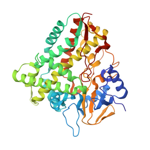
Ketoconazole-induced conformational changes in the active site of cytochrome P450eryF.

Cupp-Vickery, J.R.Garcia, C.Hofacre, A.McGee-Estrada, K.

(2001) J Mol Biology 311: 101-110

  • DOI: https://doi.org/10.1006/jmbi.2001.4803
  • Primary Citation Related Structures: 
    1JIN, 1JIO, 1JIP

  • PubMed Abstract: 

    The azole-based P450 inhibitor ketoconazole is used to treat fungal infections and functions by blocking ergosterol biosynthesis in yeast. Ketoconazole binds to mammalian P450 enzymes and this can result in drug-drug interactions and lead to liver damage. To identify protein-drug interactions that contribute to binding specificity and affinity, we determined the crystal structure of ketoconazole complexed with P450eryF. In the P450eryF/ketoconazole structure, the azole moiety and nearby rings of ketoconzole are positioned in the active site similar to the substrate, 6-deoxyerythronolide B, with the azole nitrogen atom coordinated to the heme iron atom. The remainder of the ketoconazole molecule extends into the active-site pocket, which is occupied by water in the substrate complex. Binding of ketoconazole led to unexpected conformational changes in the I-helix. The I-helix cleft near the active site has collapsed with a helical pitch of 5.4 A compared to 6.6 A in the substrate complex. P450eryF/ketoconazole crystals soaked in 6-deoxyerythronolide B to exchange ligands exhibit a structure identical with that of the original P450eryF/substrate complex, with the I-helix cleft restored to a pitch of 6.6 A. These findings indicate that the I-helix region of P450eryF is flexible and can adopt multiple conformations. An improved understanding of the flexibility of the active-site region of cytochrome P450 enzymes is important to gain insight into determinants of ligand binding/specificity as well as to evaluate models for catalytic mechanism based on static crystal structures.


  • Organizational Affiliation
    • Department of Chemistry and Biochemistry, California State University, Fullerton, 800 N. State College Blvd., Fullerton, CA 92834, USA. jvickery@fullerton.edu

Macromolecule Content 

  • Total Structure Weight: 45.66 kDa 
  • Atom Count: 3,469 
  • Modeled Residue Count: 403 
  • Deposited Residue Count: 403 
  • Unique protein chains: 1

Macromolecules

Find similar proteins by:|  3D Structure
Entity ID: 1
MoleculeChains  Sequence LengthOrganismDetailsImage
CYTOCHROME P450 107A1403Saccharopolyspora erythraeaMutation(s): 0 
EC: 1 (PDB Primary Data), 1.14.15.35 (UniProt)
UniProt
Find proteins for Q00441 (Saccharopolyspora erythraea (strain ATCC 11635 / DSM 40517 / JCM 4748 / NBRC 13426 / NCIMB 8594 / NRRL 2338))
Explore Q00441 
Go to UniProtKB:  Q00441
Entity Groups
Sequence Clusters30% Identity50% Identity70% Identity90% Identity95% Identity100% Identity
UniProt GroupQ00441
Sequence Annotations
Expand
Reference Sequence

Small Molecules

Ligands 2 Unique
IDChains Name / Formula / InChI Key2D Diagram3D Interactions
HEM

Query on HEM


Download:Ideal Coordinates CCD File
B [auth A]PROTOPORPHYRIN IX CONTAINING FE
C34 H32 Fe N4 O4
KABFMIBPWCXCRK-RGGAHWMASA-L
KTN

Query on KTN


Download:Ideal Coordinates CCD File
C [auth A]CIS-1-ACETYL-4-(4-((2-(2,4-DICHLOROPHENYL)-2-(1H-IMIDAZOL-1-YLMETHYL)-1,3-DIOXOLAN-4-YL)METHOXY)PHENYL)PIPERAZINE
C26 H28 Cl2 N4 O4
XMAYWYJOQHXEEK-JYFHCDHNSA-N

Experimental Data & Validation

Experimental Data

  • Method: X-RAY DIFFRACTION
  • Resolution: 2.30 Å
  • R-Value Free:  0.235 (Depositor), 0.189 (DCC) 
  • R-Value Work:  0.184 (Depositor) 
  • R-Value Observed: 0.184 (Depositor) 
Space Group: P 21 21 21
Unit Cell:
Length ( Å )Angle ( ˚ )
a = 54.228α = 90
b = 79.738β = 90
c = 99.364γ = 90
Software Package:
Software NamePurpose
CNSrefinement
DENZOdata reduction
SCALEPACKdata scaling
CNSphasing

Structure Validation

View Full Validation Report


Ligand Structure Quality Assessment 


Entry History 

Deposition Data

Revision History  (Full details and data files)

  • Version 1.0: 2001-10-17
    Type: Initial release
  • Version 1.1: 2008-04-27
    Changes: Version format compliance
  • Version 1.2: 2011-07-13
    Changes: Version format compliance
  • Version 1.3: 2024-02-07
    Changes: Data collection, Database references, Derived calculations
  • Version 1.4: 2024-04-03
    Changes: Refinement description