1JIB

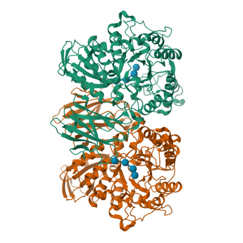
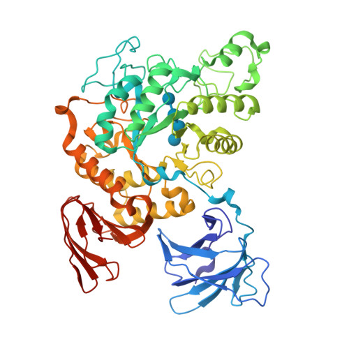
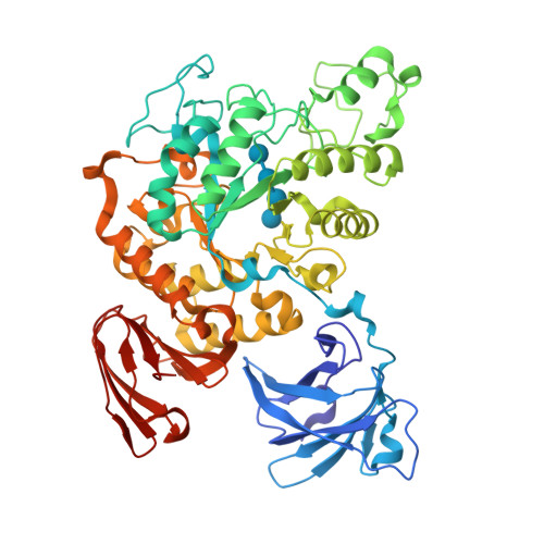
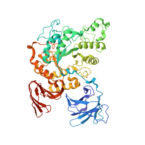
Complex of Alpha-amylase II (TVA II) from Thermoactinomyces vulgaris R-47 with Maltotetraose Based on a Crystal Soaked with Maltohexaose.


Experimental Data Snapshot

  • Method: X-RAY DIFFRACTION
  • Resolution: 3.30 Å
  • R-Value Free: 0.304 
  • R-Value Work: 0.212 

Starting Model: experimental
View more details

wwPDB Validation   3D Report Full Report


This is version 2.2 of the entry. See complete history


Literature

Structures of Thermoactinomyces vulgaris R-47 alpha-amylase II complexed with substrate analogues.

Yokota, T.Tonozuka, T.Shimura, Y.Ichikawa, K.Kamitori, S.Sakano, Y.

(2001) Biosci Biotechnol Biochem 65: 619-626

  • DOI: https://doi.org/10.1271/bbb.65.619
  • Primary Citation of Related Structures:  
    1JIB, 1JL8

  • PubMed Abstract: 

    The structures of Thermoactinomyces vulgaris R-47 alpha-amylase II mutant (d325nTVA II) complexed with substrate analogues, methyl beta-cyclodextrin (m beta-CD) and maltohexaose (G6), were solved by X-ray diffraction at 3.2 A and 3.3 A resolution, respectively. In d325nTVA II-m beta-CD complex, the orientation and binding-position of beta-CD in TVA II were identical to those in cyclodextin glucanotransferase (CGTase). The active site residues were essentialy conserved, while there are no residues corresponding to Tyr89, Phe183, and His233 of CGTase in TVA II. In d325nTVA II-G6 complex, the electron density maps of two glucosyl units at the non-reducing end were disordered and invisible. The four glucosyl units of G6 were bound to TVA II as in CGTase, while the others were not stacked and were probably flexible. The residues of TVA II corresponding to Tyr89, Lys232, and His233 of CGTase were completely lacking. These results suggest that the lack of the residues related to alpha-glucan and CD-stacking causes the functional distinctions between CGTase and TVA II.


  • Organizational Affiliation

    Department of Applied Biological Science, Tokyo University of Agriculture and Technology, Fuchu-shi, Japan.


Macromolecules
Find similar proteins by:  (by identity cutoff)  |  3D Structure
Entity ID: 1
MoleculeChains Sequence LengthOrganismDetailsImage
NEOPULLULANASE
A, B
585Thermoactinomyces vulgarisMutation(s): 1 
EC: 3.2.1.135
UniProt
Find proteins for Q08751 (Thermoactinomyces vulgaris)
Explore Q08751 
Go to UniProtKB:  Q08751
Entity Groups  
Sequence Clusters30% Identity50% Identity70% Identity90% Identity95% Identity100% Identity
UniProt GroupQ08751
Sequence Annotations
Expand
  • Reference Sequence
Oligosaccharides

Help

Entity ID: 2
MoleculeChains Length2D Diagram Glycosylation3D Interactions
alpha-D-glucopyranose-(1-4)-alpha-D-glucopyranose-(1-4)-alpha-D-glucopyranose-(1-4)-alpha-D-glucopyranose
C, D
4N/A
Glycosylation Resources
GlyTouCan:  G87171PZ
GlyCosmos:  G87171PZ
GlyGen:  G87171PZ
Biologically Interesting Molecules (External Reference) 1 Unique
Experimental Data & Validation

Experimental Data

  • Method: X-RAY DIFFRACTION
  • Resolution: 3.30 Å
  • R-Value Free: 0.304 
  • R-Value Work: 0.212 
  • Space Group: P 21 21 21
Unit Cell:
Length ( Å )Angle ( ˚ )
a = 111.87α = 90
b = 117.32β = 90
c = 112.61γ = 90
Software Package:
Software NamePurpose
CNSrefinement
MOSFLMdata reduction
CCP4data scaling
CNSphasing

Structure Validation

View Full Validation Report


Entry History 

Deposition Data

Revision History  (Full details and data files)

  • Version 1.0: 2001-07-25
    Type: Initial release
  • Version 1.1: 2008-04-27
    Changes: Version format compliance
  • Version 1.2: 2011-07-13
    Changes: Version format compliance
  • Version 2.0: 2020-07-29
    Type: Remediation
    Reason: Carbohydrate remediation
    Changes: Atomic model, Data collection, Derived calculations, Non-polymer description, Structure summary
  • Version 2.1: 2021-11-10
    Changes: Database references, Structure summary
  • Version 2.2: 2023-10-25
    Changes: Data collection, Refinement description