1JI3 | pdb_00001ji3

CRYSTAL STRUCTURE OF THE FIRST THERMOSTABLE BACTERIAL LIPASE FROM BACILLUS STEAROTHERMOPHILUS


Experimental Data Snapshot

  • Method: X-RAY DIFFRACTION
  • Resolution: 2.20 Å
  • R-Value Free: 
    0.210 (Depositor) 
  • R-Value Work: 
    0.166 (Depositor) 
  • R-Value Observed: 
    0.169 (Depositor) 

wwPDB Validation 3D Report Full Report

Validation slider image for 1JI3

This is version 1.4 of the entry. See complete history

Literature

Crystal Structure of a Thermostable Lipase from Bacillus stearothermophilus P1

Tyndall, J.D.A.Sinchaikul, S.Fothergill-Gilmore, L.A.Taylor, P.Walkinshaw, M.D.

(2002) J Mol Biology 323: 859-869

  • DOI: https://doi.org/10.1016/s0022-2836(02)01004-5
  • Primary Citation Related Structures: 
    1JI3

  • PubMed Abstract: 

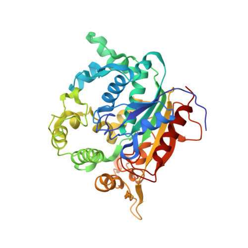
    We describe the first lipase structure from a thermophilic organism. It shares less than 20% amino acid sequence identity with other lipases for which there are crystal structures, and shows significant insertions compared with the typical alpha/beta hydrolase canonical fold. The structure contains a zinc-binding site which is unique among all lipases with known structures, and which may play a role in enhancing thermal stability. Zinc binding is mediated by two histidine and two aspartic acid residues. These residues are present in comparable positions in the sequences of certain lipases for which there is as yet no crystal structural information, such as those from Staphylococcal species and Arabidopsis thaliana. The structure of Bacillus stearothermophilus P1 lipase provides a template for other thermostable lipases, and offers insight into mechanisms used to enhance thermal stability which may be of commercial value in engineering lipases for industrial uses.


  • Organizational Affiliation
    • Structural Biochemistry Group, Institute of Cell and Molecular Biology, University of Edinburgh, Michael Swann Building, King's Buildings, Mayfield Road, Scotland, UK.

Macromolecule Content 

  • Total Structure Weight: 86.83 kDa 
  • Atom Count: 6,470 
  • Modeled Residue Count: 776 
  • Deposited Residue Count: 776 
  • Unique protein chains: 1

Macromolecules

Find similar proteins by:|  3D Structure
Entity ID: 1
MoleculeChains  Sequence LengthOrganismDetailsImage
lipase
A, B
388Geobacillus stearothermophilusMutation(s): 0 
EC: 3.1.1.3
UniProt
Find proteins for Q9L6D3 (Geobacillus stearothermophilus)
Explore Q9L6D3 
Go to UniProtKB:  Q9L6D3
Entity Groups
Sequence Clusters30% Identity50% Identity70% Identity90% Identity95% Identity100% Identity
UniProt GroupQ9L6D3
Sequence Annotations
Expand
Reference Sequence

Experimental Data & Validation

Experimental Data

  • Method: X-RAY DIFFRACTION
  • Resolution: 2.20 Å
  • R-Value Free:  0.210 (Depositor) 
  • R-Value Work:  0.166 (Depositor) 
  • R-Value Observed: 0.169 (Depositor) 
Space Group: C 1 2 1
Unit Cell:
Length ( Å )Angle ( ˚ )
a = 118.5α = 90
b = 81.237β = 96.33
c = 99.782γ = 90
Software Package:
Software NamePurpose
MAR345data collection
SCALEPACKdata scaling
SOLVEphasing
REFMACrefinement

Structure Validation

View Full Validation Report


Entry History 

Deposition Data

Revision History  (Full details and data files)

  • Version 1.0: 2002-11-01
    Type: Initial release
  • Version 1.1: 2008-04-27
    Changes: Version format compliance
  • Version 1.2: 2011-07-13
    Changes: Version format compliance
  • Version 1.3: 2017-10-04
    Changes: Refinement description
  • Version 1.4: 2024-02-07
    Changes: Data collection, Database references, Derived calculations