1JI1 | pdb_00001ji1

Crystal Structure Analysis of Thermoactinomyces vulgaris R-47 alpha-Amylase 1


Experimental Data Snapshot

  • Method: X-RAY DIFFRACTION
  • Resolution: 1.60 Å
  • R-Value Free: 
    0.206 (Depositor), 0.206 (DCC) 
  • R-Value Work: 
    0.182 (Depositor), 0.182 (DCC) 

wwPDB Validation 3D Report Full Report

Validation slider image for 1JI1

This is version 1.4 of the entry. See complete history

Literature

Crystal structures and structural comparison of Thermoactinomyces vulgaris R-47 alpha-amylase 1 (TVAI) at 1.6 A resolution and alpha-amylase 2 (TVAII) at 2.3 A resolution.

Kamitori, S.Abe, A.Ohtaki, A.Kaji, A.Tonozuka, T.Sakano, Y.

(2002) J Mol Biology 318: 443-453

  • DOI: https://doi.org/10.1016/S0022-2836(02)00111-0
  • Primary Citation Related Structures: 
    1JI1, 1JI2

  • PubMed Abstract: 

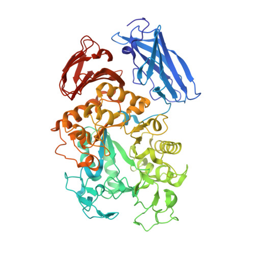
    The X-ray crystal structures of Thermoactinomyces vulgaris R-47 alpha-amylase 1 (TVAI) and alpha-amylase 2 (TVAII) have been determined at 1.6 A and 2.3 A resolution, respectively. The structures of TVAI and TVAII have been refined, R-factor of 0.182 (R(free)=0.206) and 0.179 (0.224), respectively, with good chemical geometries. Both TVAI and TVAII have four domains, N, A, B and C, and all very similar in structure. However, there are some differences in the structures between them. Domain N of TVAI interacts strongly with domains A and B, giving a spherical shape structure to the enzyme, while domain N of TVAII is isolated from the other domains, which leads to the formation of a dimer. TVAI has three bound Ca ions, whereas TVAII has only one. TVAI has eight extra loops compared to TVAII, while TVAII has two extra loops compared to TVAI. TVAI can hydrolyze substrates more efficiently than TVAII with a high molecular mass such as starch, while TVAII is much more active against cyclodextrins than TVAI and other alpha-amylases. A structural comparison of the active sites has clearly revealed this difference in substrate specificity.


  • Organizational Affiliation
    • Department of Biotechnology and Life Science, Tokyo University of Agriculture and Technology, 2-24-16 Naka-cho, Koganei, Tokyo 184-8588, Japan. kamitori@cc.tuat.ac.jp

Macromolecule Content 

  • Total Structure Weight: 142.46 kDa 
  • Atom Count: 11,556 
  • Modeled Residue Count: 1,274 
  • Deposited Residue Count: 1,274 
  • Unique protein chains: 1

Macromolecules

Find similar proteins by:|  3D Structure
Entity ID: 1
MoleculeChains  Sequence LengthOrganismDetailsImage
ALPHA-AMYLASE I
A, B
637Thermoactinomyces vulgarisMutation(s): 0 
EC: 3.2.1.1 (PDB Primary Data), 3.2.1.135 (UniProt)
UniProt
Find proteins for Q60053 (Thermoactinomyces vulgaris)
Explore Q60053 
Go to UniProtKB:  Q60053
Entity Groups
Sequence Clusters30% Identity50% Identity70% Identity90% Identity95% Identity100% Identity
UniProt GroupQ60053
Sequence Annotations
Expand
Reference Sequence

Experimental Data & Validation

Experimental Data

  • Method: X-RAY DIFFRACTION
  • Resolution: 1.60 Å
  • R-Value Free:  0.206 (Depositor), 0.206 (DCC) 
  • R-Value Work:  0.182 (Depositor), 0.182 (DCC) 
Space Group: P 1 21 1
Unit Cell:
Length ( Å )Angle ( ˚ )
a = 122.57α = 90
b = 50.84β = 104.2
c = 107.8γ = 90
Software Package:
Software NamePurpose
MLPHAREphasing
CNSrefinement
MOSFLMdata reduction
CCP4data scaling

Structure Validation

View Full Validation Report


Entry History 

Deposition Data

Revision History  (Full details and data files)

  • Version 1.0: 2002-06-05
    Type: Initial release
  • Version 1.1: 2008-04-27
    Changes: Version format compliance
  • Version 1.2: 2011-07-13
    Changes: Version format compliance
  • Version 1.3: 2014-04-16
    Changes: Other
  • Version 1.4: 2024-03-13
    Changes: Data collection, Database references, Derived calculations