1JGM | pdb_00001jgm

High Resolution Structure of the Cadmium-containing Phosphotriesterase from Pseudomonas diminuta


Experimental Data Snapshot

  • Method: X-RAY DIFFRACTION
  • Resolution: 1.30 Å
  • R-Value Free: 
    0.236 (Depositor) 
  • R-Value Work: 
    0.202 (Depositor) 
  • R-Value Observed: 
    0.204 (Depositor) 

wwPDB Validation 3D Report Full Report

Validation slider image for 1JGM

This is version 1.3 of the entry. See complete history

Literature

High resolution X-ray structures of different metal-substituted forms of phosphotriesterase from Pseudomonas diminuta.

Benning, M.M.Shim, H.Raushel, F.M.Holden, H.M.

(2001) Biochemistry 40: 2712-2722

  • DOI: https://doi.org/10.1021/bi002661e
  • Primary Citation Related Structures: 
    1HZY, 1I0B, 1I0D, 1JGM

  • PubMed Abstract: 

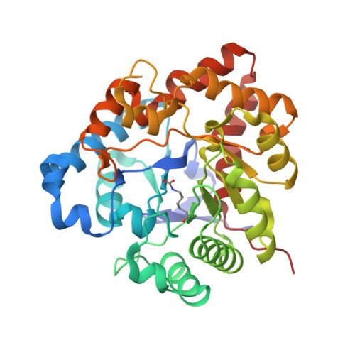
    Phosphotriesterase, isolated from the soil-dwelling bacterium Pseudomonas diminuta, catalyzes the detoxification of organophosphate-based insecticides and chemical warfare agents. The enzyme has attracted significant research attention in light of its possible employment as a bioremediation tool. As naturally isolated, the enzyme is dimeric. Each subunit contains a binuclear zinc center that is situated at the C-terminal portion of a "TIM" barrel motif. The two zincs are separated by approximately 3.4 A and coordinated to the protein via the side chains of His 55, His 57, His 201, His 230, Asp 301, and a carboxylated Lys 169. Both Lys 169 and a water molecule (or hydroxide ion) serve to bridge the two zinc ions together. Interestingly, these metals can be replaced with cadmium or manganese ions without loss of enzymatic activity. Here we describe the three-dimensional structures of the Zn(2+)/Zn(2+)-, Zn(2+)/Cd(2+)-, Cd(2+)/Cd(2+)-, and Mn(2+)/Mn(2+)-substituted forms of phosphotriesterase determined and refined to a nominal resolution of 1.3 A. In each case, the more buried metal ion, referred to as the alpha-metal, is surrounded by ligands in a trigonal bipyramidal ligation sphere. For the more solvent-exposed or beta-metal ion, however, the observed coordination spheres are either octahedral (in the Cd(2+)/Cd(2+)-, Mn(2+)/Mn(2+)-, and the mixed Zn(2+)/Cd(2+)-species) or trigonal bipyramidal (in the Zn(2+)/Zn(2+)-protein). By measuring the anomalous X-ray data from crystals of the Zn(2+)/Cd(2+)-species, it has been possible to determine that the alpha-metal ion is zinc and the beta-site is occupied by cadmium.


  • Organizational Affiliation
    • Department of Biochemistry, University of Wisconsin, Madison, Wisconsin 53706, USA.

Macromolecule Content 

  • Total Structure Weight: 75.29 kDa 
  • Atom Count: 5,997 
  • Modeled Residue Count: 665 
  • Deposited Residue Count: 672 
  • Unique protein chains: 1

Macromolecules

Find similar proteins by:|  3D Structure
Entity ID: 1
MoleculeChains  Sequence LengthOrganismDetailsImage
Phosphotriesterase
A, B
336Brevundimonas diminutaMutation(s): 1 
Gene Names: OPD
EC: 3.1.8.1
UniProt
Find proteins for P0A434 (Brevundimonas diminuta)
Explore P0A434 
Go to UniProtKB:  P0A434
Entity Groups
Sequence Clusters30% Identity50% Identity70% Identity90% Identity95% Identity100% Identity
UniProt GroupP0A434
Sequence Annotations
Expand
Reference Sequence

Small Molecules

Ligands 4 Unique
IDChains Name / Formula / InChI Key2D Diagram3D Interactions
PEL

Query on PEL


Download:Ideal Coordinates CCD File
LA [auth B],
U [auth A],
V [auth A]
2-PHENYL-ETHANOL
C8 H10 O
WRMNZCZEMHIOCP-UHFFFAOYSA-N
CD

Query on CD


Download:Ideal Coordinates CCD File
C [auth A],
D [auth A],
W [auth B],
X [auth B]
CADMIUM ION
Cd
WLZRMCYVCSSEQC-UHFFFAOYSA-N
EDO

Query on EDO


Download:Ideal Coordinates CCD File
AA [auth B]
BA [auth B]
CA [auth B]
DA [auth B]
EA [auth B]
AA [auth B],
BA [auth B],
CA [auth B],
DA [auth B],
EA [auth B],
F [auth A],
FA [auth B],
G [auth A],
GA [auth B],
H [auth A],
HA [auth B],
I [auth A],
IA [auth B],
J [auth A],
JA [auth B],
K [auth A],
KA [auth B],
L [auth A],
M [auth A],
N [auth A],
O [auth A],
P [auth A],
Q [auth A],
R [auth A],
S [auth A],
T [auth A],
Z [auth B]
1,2-ETHANEDIOL
C2 H6 O2
LYCAIKOWRPUZTN-UHFFFAOYSA-N
NA

Query on NA


Download:Ideal Coordinates CCD File
E [auth A],
Y [auth B]
SODIUM ION
Na
FKNQFGJONOIPTF-UHFFFAOYSA-N
Modified Residues  1 Unique
IDChains TypeFormula2D DiagramParent
KCX
Query on KCX
A, B
L-PEPTIDE LINKINGC7 H14 N2 O4LYS

Experimental Data & Validation

Experimental Data

  • Method: X-RAY DIFFRACTION
  • Resolution: 1.30 Å
  • R-Value Free:  0.236 (Depositor) 
  • R-Value Work:  0.202 (Depositor) 
  • R-Value Observed: 0.204 (Depositor) 
Space Group: C 1 2 1
Unit Cell:
Length ( Å )Angle ( ˚ )
a = 128.45α = 90
b = 90.03β = 91.72
c = 68.39γ = 90
Software Package:
Software NamePurpose
TNTrefinement
d*TREKdata reduction
HKL-2000data scaling
TNTphasing

Structure Validation

View Full Validation Report


Entry History 

Deposition Data

Revision History  (Full details and data files)

  • Version 1.0: 2001-07-04
    Type: Initial release
  • Version 1.1: 2008-04-27
    Changes: Version format compliance
  • Version 1.2: 2011-07-13
    Changes: Version format compliance
  • Version 1.3: 2025-03-26
    Changes: Data collection, Database references, Derived calculations, Structure summary