1JGJ | pdb_00001jgj

CRYSTAL STRUCTURE OF SENSORY RHODOPSIN II AT 2.4 ANGSTROMS: INSIGHTS INTO COLOR TUNING AND TRANSDUCER INTERACTION


Experimental Data Snapshot

  • Method: X-RAY DIFFRACTION
  • Resolution: 2.40 Å
  • R-Value Free: 
    0.280 (Depositor) 
  • R-Value Work: 
    0.233 (Depositor) 
  • R-Value Observed: 
    0.233 (Depositor) 

Starting Model: experimental
View more details

wwPDB Validation 3D Report Full Report

Validation slider image for 1JGJ

This is version 1.5 of the entry. See complete history

Literature

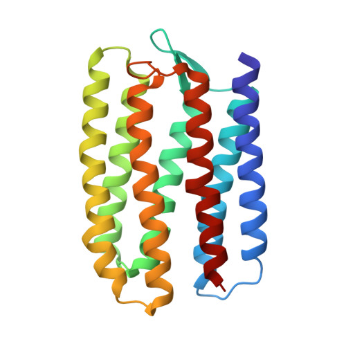
Crystal structure of sensory rhodopsin II at 2.4 angstroms: insights into color tuning and transducer interaction.

Luecke, H.Schobert, B.Lanyi, J.K.Spudich, E.N.Spudich, J.L.

(2001) Science 293: 1499-1503

  • DOI: https://doi.org/10.1126/science.1062977
  • Primary Citation Related Structures: 
    1JGJ

  • PubMed Abstract: 

    We report an atomic-resolution structure for a sensory member of the microbial rhodopsin family, the phototaxis receptor sensory rhodopsin II (NpSRII), which mediates blue-light avoidance by the haloarchaeon Natronobacterium pharaonis. The 2.4 angstrom structure reveals features responsible for the 70- to 80-nanometer blue shift of its absorption maximum relative to those of haloarchaeal transport rhodopsins, as well as structural differences due to its sensory, as opposed to transport, function. Multiple factors appear to account for the spectral tuning difference with respect to bacteriorhodopsin: (i) repositioning of the guanidinium group of arginine 72, a residue that interacts with the counterion to the retinylidene protonated Schiff base; (ii) rearrangement of the protein near the retinal ring; and (iii) changes in tilt and slant of the retinal polyene chain. Inspection of the surface topography reveals an exposed polar residue, tyrosine 199, not present in bacteriorhodopsin, in the middle of the membrane bilayer. We propose that this residue interacts with the adjacent helices of the cognate NpSRII transducer NpHtrII.


  • Organizational Affiliation
    • Department of Molecular Biology and Biochemistry, University of California, Irvine, CA 92697, USA. hudel@uci.edu

Macromolecule Content 

  • Total Structure Weight: 23.75 kDa 
  • Atom Count: 1,683 
  • Modeled Residue Count: 217 
  • Deposited Residue Count: 217 
  • Unique protein chains: 1

Macromolecules

Find similar proteins by:|  3D Structure
Entity ID: 1
MoleculeChains  Sequence LengthOrganismDetailsImage
SENSORY RHODOPSIN II217Natronomonas pharaonisMutation(s): 0 
Membrane Entity: Yes 
UniProt
Find proteins for P42196 (Natronomonas pharaonis)
Explore P42196 
Go to UniProtKB:  P42196
Entity Groups
Sequence Clusters30% Identity50% Identity70% Identity90% Identity95% Identity100% Identity
UniProt GroupP42196
Sequence Annotations
Expand
Reference Sequence

Small Molecules

Ligands 2 Unique
IDChains Name / Formula / InChI Key2D Diagram3D Interactions
BOG

Query on BOG


Download:Ideal Coordinates CCD File
B [auth A]octyl beta-D-glucopyranoside
C14 H28 O6
HEGSGKPQLMEBJL-RKQHYHRCSA-N
RET

Query on RET


Download:Ideal Coordinates CCD File
C [auth A]RETINAL
C20 H28 O
NCYCYZXNIZJOKI-OVSJKPMPSA-N

Experimental Data & Validation

Experimental Data

  • Method: X-RAY DIFFRACTION
  • Resolution: 2.40 Å
  • R-Value Free:  0.280 (Depositor) 
  • R-Value Work:  0.233 (Depositor) 
  • R-Value Observed: 0.233 (Depositor) 
Space Group: C 2 2 21
Unit Cell:
Length ( Å )Angle ( ˚ )
a = 87.34α = 90
b = 130.81β = 90
c = 50.87γ = 90
Software Package:
Software NamePurpose
X-PLORmodel building
CNSrefinement
DENZOdata reduction
SCALEPACKdata scaling
X-PLORphasing

Structure Validation

View Full Validation Report


Entry History 

Deposition Data

  • Released Date: 2001-07-18 
  • Deposition Author(s): Luecke, H.

Revision History  (Full details and data files)

  • Version 1.0: 2001-07-18
    Type: Initial release
  • Version 1.1: 2008-04-27
    Changes: Version format compliance
  • Version 1.2: 2011-07-13
    Changes: Version format compliance
  • Version 1.3: 2020-07-29
    Type: Remediation
    Reason: Carbohydrate remediation
    Changes: Data collection, Database references, Derived calculations, Structure summary
  • Version 1.4: 2023-08-16
    Changes: Data collection, Database references, Refinement description, Structure summary
  • Version 1.5: 2024-10-16
    Changes: Structure summary