1JF9 | pdb_00001jf9

Crystal Structure of selenocysteine lyase


Experimental Data Snapshot

  • Method: X-RAY DIFFRACTION
  • Resolution: 2.00 Å
  • R-Value Free: 
    0.207 (Depositor), 0.175 (DCC) 
  • R-Value Work: 
    0.178 (Depositor) 

wwPDB Validation 3D Report Full Report

Validation slider image for 1JF9

Ligand Structure Quality Assessment 


This is version 1.5 of the entry. See complete history

Literature

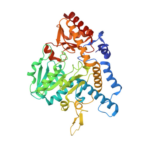
Analysis of the E. coli NifS CsdB Protein at 2.0A Reveals the Structural Basis for Perselenide and Persulfide Intermediate Formation

Lima, C.D.

(2002) J Mol Biology 315: 1199-1208

  • DOI: https://doi.org/10.1006/jmbi.2001.5308
  • Primary Citation Related Structures: 
    1JF9, 1KMJ, 1KMK

  • PubMed Abstract: 

    The Escherichia coli NifS CsdB protein is a member of the homodimeric pyridoxal 5'-phosphate (PLP)-dependent family of enzymes. These enzymes are capable of decomposing cysteine or selenocysteine into L-alanine and sulfur or selenium, respectively. E. coli NifS CsdB has a high specificity for L-selenocysteine in comparison to l-cysteine, suggesting a role for this enzyme is selenium metabolism. The 2.0 A crystal structure of E. coli NifS CsdB reveals a high-resolution view of the active site of this enzyme in apo-, persulfide, perselenide, and selenocysteine-bound intermediates, suggesting a mechanism for the stabilization of the enzyme persulfide and perselenide intermediates during catalysis, a necessary intermediate in the formation of sulfur and selenium containing metabolites.


  • Organizational Affiliation
    • Biochemistry Department and Structural Biology Program, Weill Medical College of Cornell University, New York, NY 10021, USA. lima@pinky.med.cornell.edu

Macromolecule Content 

  • Total Structure Weight: 44.87 kDa 
  • Atom Count: 3,713 
  • Modeled Residue Count: 405 
  • Deposited Residue Count: 408 
  • Unique protein chains: 1

Macromolecules

Find similar proteins by:|  3D Structure
Entity ID: 1
MoleculeChains  Sequence LengthOrganismDetailsImage
SELENOCYSTEINE LYASE408Escherichia coliMutation(s): 0 
Gene Names: NIFS/CSDB
EC: 4.4.1.16 (PDB Primary Data), 3.13.1 (UniProt), 2.8.1.7 (UniProt)
UniProt
Find proteins for P77444 (Escherichia coli (strain K12))
Explore P77444 
Go to UniProtKB:  P77444
Entity Groups
Sequence Clusters30% Identity50% Identity70% Identity90% Identity95% Identity100% Identity
UniProt GroupP77444
Sequence Annotations
Expand
Reference Sequence

Small Molecules

Ligands 1 Unique
IDChains Name / Formula / InChI Key2D Diagram3D Interactions
PLP

Query on PLP


Download:Ideal Coordinates CCD File
B [auth A]PYRIDOXAL-5'-PHOSPHATE
C8 H10 N O6 P
NGVDGCNFYWLIFO-UHFFFAOYSA-N

Experimental Data & Validation

Experimental Data

  • Method: X-RAY DIFFRACTION
  • Resolution: 2.00 Å
  • R-Value Free:  0.207 (Depositor), 0.175 (DCC) 
  • R-Value Work:  0.178 (Depositor) 
Space Group: P 43 21 2
Unit Cell:
Length ( Å )Angle ( ˚ )
a = 126.375α = 90
b = 126.375β = 90
c = 133.578γ = 90
Software Package:
Software NamePurpose
DENZOdata reduction
SCALEPACKdata scaling
SOLVEphasing
REFMACrefinement

Structure Validation

View Full Validation Report


Ligand Structure Quality Assessment 


Entry History 

Deposition Data

Revision History  (Full details and data files)

  • Version 1.0: 2001-07-04
    Type: Initial release
  • Version 1.1: 2008-04-27
    Changes: Version format compliance
  • Version 1.2: 2011-07-13
    Changes: Derived calculations, Version format compliance
  • Version 1.3: 2015-01-21
    Changes: Refinement description
  • Version 1.4: 2021-02-03
    Changes: Database references, Derived calculations, Structure summary
  • Version 1.5: 2025-03-26
    Changes: Data collection, Database references, Structure summary