1JEH | pdb_00001jeh

CRYSTAL STRUCTURE OF YEAST E3, LIPOAMIDE DEHYDROGENASE


Experimental Data Snapshot

  • Method: X-RAY DIFFRACTION
  • Resolution: 2.40 Å
  • R-Value Free: 
    0.260 (Depositor) 
  • R-Value Work: 
    0.202 (Depositor) 
  • R-Value Observed: 
    0.202 (Depositor) 

Starting Model: experimental
View more details

wwPDB Validation 3D Report Full Report

Validation slider image for 1JEH

This is version 1.7 of the entry. See complete history

Literature

Crystal structure of eucaryotic E3, lipoamide dehydrogenase from yeast.

Toyoda, T.Suzuki, K.Sekiguchi, T.Reed, L.J.Takenaka, A.

(1998) J Biochem 123: 668-674

  • DOI: https://doi.org/10.1093/oxfordjournals.jbchem.a021989
  • Primary Citation Related Structures: 
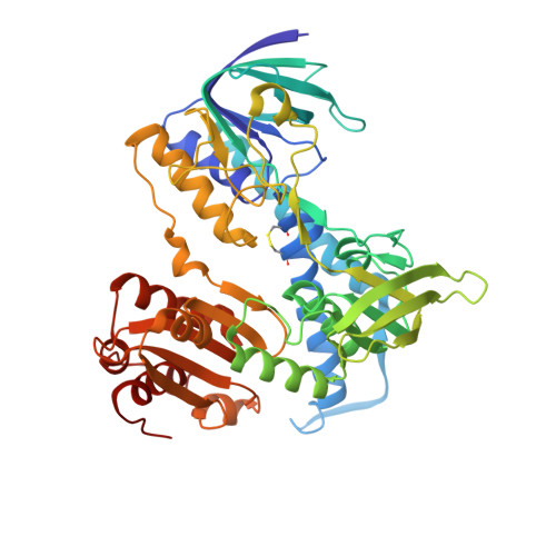
    1JEH

  • PubMed Abstract: 

    The crystal structure of eucaryotic lipoamide dehydrogenase from yeast has been determined by an X-ray analysis at 2.7 (partially at 2.4) A resolution. The enzyme has two identical subunits related by a pseudo twofold symmetry. The tertiary structure is similar to those of other procaryotic enzymes. The active site, consisting of FAD, Cys44, and Cys49 from one subunit and His457' from the other subunit, is highly conserved. This enzyme is directly bound to the core protein E2 of the 2-oxoglutarate dehydrogenase complex, whereas it is bound to the pyruvate dehydrogenase complex through a protein X. The calculated electrostatic potential suggests two characteristic regions for binding with these two proteins.


  • Organizational Affiliation
    • Department of Life Science, Faculty of Bioscience and Biotechnology, Tokyo Institute of Technology, Yokohama 226-8501.

Macromolecule Content 

  • Total Structure Weight: 104.82 kDa 
  • Atom Count: 7,363 
  • Modeled Residue Count: 956 
  • Deposited Residue Count: 956 
  • Unique protein chains: 1

Macromolecules

Find similar proteins by:|  3D Structure
Entity ID: 1
MoleculeChains  Sequence LengthOrganismDetailsImage
DIHYDROLIPOAMIDE DEHYDROGENASE
A, B
478Saccharomyces cerevisiaeMutation(s): 0 
EC: 1.8.1.4
UniProt
Find proteins for P09624 (Saccharomyces cerevisiae (strain ATCC 204508 / S288c))
Explore P09624 
Go to UniProtKB:  P09624
Entity Groups
Sequence Clusters30% Identity50% Identity70% Identity90% Identity95% Identity100% Identity
UniProt GroupP09624
Sequence Annotations
Expand
Reference Sequence

Small Molecules

Ligands 1 Unique
IDChains Name / Formula / InChI Key2D Diagram3D Interactions
FAD

Query on FAD


Download:Ideal Coordinates CCD File
C [auth A],
D [auth B]
FLAVIN-ADENINE DINUCLEOTIDE
C27 H33 N9 O15 P2
VWWQXMAJTJZDQX-UYBVJOGSSA-N

Experimental Data & Validation

Experimental Data

  • Method: X-RAY DIFFRACTION
  • Resolution: 2.40 Å
  • R-Value Free:  0.260 (Depositor) 
  • R-Value Work:  0.202 (Depositor) 
  • R-Value Observed: 0.202 (Depositor) 
Space Group: P 21 21 21
Unit Cell:
Length ( Å )Angle ( ˚ )
a = 97.1α = 90
b = 158.7β = 90
c = 67.9γ = 90
Software Package:
Software NamePurpose
DENZOdata reduction
ROTAVATAdata reduction
MERLOTphasing
X-PLORrefinement
CCP4data scaling
ROTAVATAdata scaling

Structure Validation

View Full Validation Report


Entry History 

Deposition Data

Revision History  (Full details and data files)

  • Version 1.0: 2001-07-11
    Type: Initial release
  • Version 1.1: 2008-04-27
    Changes: Version format compliance
  • Version 1.2: 2011-07-13
    Changes: Version format compliance
  • Version 1.3: 2017-10-04
    Changes: Advisory, Refinement description
  • Version 1.4: 2018-02-07
    Changes: Experimental preparation
  • Version 1.5: 2019-11-06
    Changes: Data collection, Database references
  • Version 1.6: 2023-10-25
    Changes: Advisory, Data collection, Database references, Derived calculations, Refinement description
  • Version 1.7: 2024-11-20
    Changes: Structure summary