1JEF

TURKEY LYSOZYME COMPLEX WITH (GLCNAC)3


Experimental Data Snapshot

  • Method: X-RAY DIFFRACTION
  • Resolution: 1.77 Å
  • R-Value Free: 0.225 
  • R-Value Work: 0.182 
  • R-Value Observed: 0.182 

Starting Model: experimental
View more details

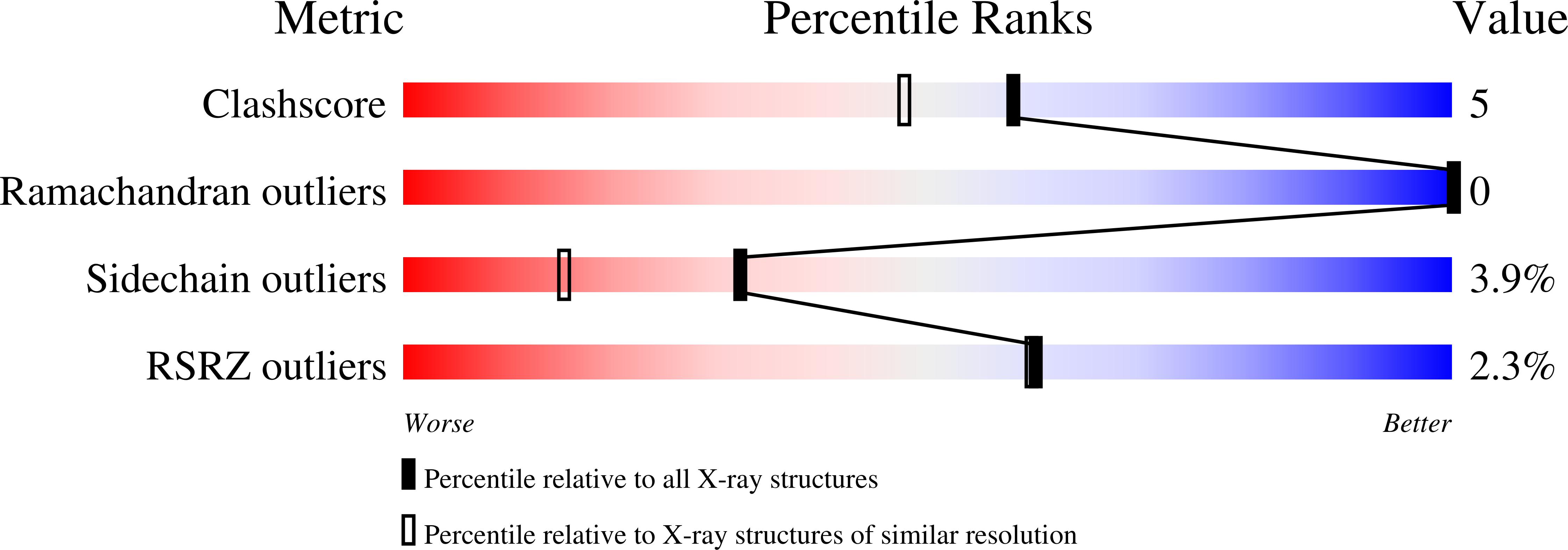
wwPDB Validation   3D Report Full Report


This is version 2.2 of the entry. See complete history


Literature

X-ray structure of turkey-egg lysozyme complex with tri-N-acetylchitotriose. Lack of binding ability at subsite A.

Harata, K.Muraki, M.

(1997) Acta Crystallogr D Biol Crystallogr 53: 650-657

  • DOI: https://doi.org/10.1107/S0907444997005362
  • Primary Citation of Related Structures:  
    1JEF

  • PubMed Abstract: 

    The turkey-egg lysozyme (TEL) complex with tri-N-acetylchitotriose [(GlcNac)3] was co-crystallized from 1.5% TEL and 2 mM (GlcNac)3 at pH 4.2. The crystal structure was determined by molecular replacement and refined to an R value of 0.182 using 10-1.77 A data. The (GlcNac)3 molecule occupies the subsites A, B and C. At the subsites B and C, the sugar residues are bound in a similar manner to that found in the hen-egg lysozyme (HEL) complex. In contrast, the GlcNac residue at the subsite A is exposed to bulk solvent and has no contact with the protein molecule because the active residue Asp101 in HEL is replaced by Gly in TEL. A sulfate ion is bound in the vicinity of subsite B and forms hydrogen bonds with the sugar residue and the guanidino group of Arg61, assisting the binding of the sugar residue to subsite B. The active-site cleft of TEL is narrower than that of native TEL, thus attaining the best fit of the (GlcNac)3 molecule. The lack of binding ability of subsite A is discussed in relation to the catalytic properties of TEL. The result suggests that the cleavage pattern of oligosaccharide substrates in the catalytic reaction is regulated by the protein-sugar interaction at subsite A.


  • Organizational Affiliation

    National Institute of Bioscience and Human Technology, Tsukuba, Ibaraki, Japan. harata@nibh.go.jp


Macromolecules
Find similar proteins by:  (by identity cutoff)  |  3D Structure
Entity ID: 1
MoleculeChains Sequence LengthOrganismDetailsImage
LYSOZYME129Meleagris gallopavoMutation(s): 0 
EC: 3.2.1.17
UniProt
Find proteins for P00703 (Meleagris gallopavo)
Explore P00703 
Go to UniProtKB:  P00703
Entity Groups  
Sequence Clusters30% Identity50% Identity70% Identity90% Identity95% Identity100% Identity
UniProt GroupP00703
Sequence Annotations
Expand
  • Reference Sequence
Oligosaccharides

Help

Entity ID: 2
MoleculeChains Length2D Diagram Glycosylation3D Interactions
2-acetamido-2-deoxy-beta-D-glucopyranose-(1-4)-2-acetamido-2-deoxy-beta-D-glucopyranose-(1-4)-2-acetamido-2-deoxy-beta-D-glucopyranose
B
3N/A
Glycosylation Resources
GlyTouCan:  G47362BJ
GlyCosmos:  G47362BJ
GlyGen:  G47362BJ
Small Molecules
Ligands 1 Unique
IDChains Name / Formula / InChI Key2D Diagram3D Interactions
SO4
Query on SO4

Download Ideal Coordinates CCD File 
C [auth A]SULFATE ION
O4 S
QAOWNCQODCNURD-UHFFFAOYSA-L
Biologically Interesting Molecules (External Reference) 1 Unique
Experimental Data & Validation

Experimental Data

  • Method: X-RAY DIFFRACTION
  • Resolution: 1.77 Å
  • R-Value Free: 0.225 
  • R-Value Work: 0.182 
  • R-Value Observed: 0.182 
  • Space Group: C 1 2 1
Unit Cell:
Length ( Å )Angle ( ˚ )
a = 87.49α = 90
b = 32.94β = 119.1
c = 49γ = 90
Software Package:
Software NamePurpose
MADNESdata collection
MERGEFdata reduction
X-PLORmodel building
X-PLORrefinement
MADNESdata reduction
MERGEFdata scaling
X-PLORphasing

Structure Validation

View Full Validation Report


Entry History 

Deposition Data

Revision History  (Full details and data files)

  • Version 1.0: 1997-10-15
    Type: Initial release
  • Version 1.1: 2008-03-24
    Changes: Version format compliance
  • Version 1.2: 2011-07-13
    Changes: Non-polymer description, Version format compliance
  • Version 1.3: 2018-04-18
    Changes: Data collection, Other
  • Version 2.0: 2020-07-29
    Type: Remediation
    Reason: Carbohydrate remediation
    Changes: Atomic model, Data collection, Derived calculations, Structure summary
  • Version 2.1: 2024-04-03
    Changes: Data collection, Database references, Refinement description, Structure summary
  • Version 2.2: 2024-10-16
    Changes: Structure summary