1JEE | pdb_00001jee

Crystal Structure of ATP Sulfurylase in complex with chlorate


Experimental Data Snapshot

  • Method: X-RAY DIFFRACTION
  • Resolution: 2.80 Å
  • R-Value Free: 
    0.232 (Depositor), 0.231 (DCC) 
  • R-Value Work: 
    0.184 (Depositor), 0.184 (DCC) 
  • R-Value Observed: 
    0.184 (Depositor) 

Starting Model: experimental
View more details

wwPDB Validation 3D Report Full Report

Validation slider image for 1JEE

Ligand Structure Quality Assessment 


This is version 1.3 of the entry. See complete history

Literature

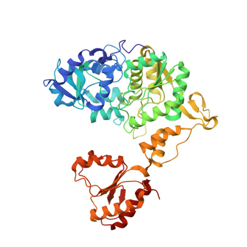
The complex structures of ATP sulfurylase with thiosulfate, ADP and chlorate reveal new insights in inhibitory effects and the catalytic cycle.

Ullrich, T.C.Huber, R.

(2001) J Mol Biology 313: 1117-1125

  • DOI: https://doi.org/10.1006/jmbi.2001.5098
  • Primary Citation Related Structures: 
    1JEC, 1JED, 1JEE

  • PubMed Abstract: 

    The ubiquitous enzyme ATP sulfurylase (ATPS) catalyzes the primary step of intracellular sulfate activation, the formation of adenosine 5'-phosphosulfate (APS). It has been shown that the enzyme catalyzes the generation of APS from ATP and inorganic sulfate in vitro and in vivo, and that this reaction can be inhibited by a number of simple molecules. Here, we present the crystal structures of ATPS from the yeast Saccharomyces cerevisiae complexed with compounds that have inhibitory effects on the catalytic reaction of ATPS. Thiosulfate and ADP mimic the substrates sulfate and ATP in the active site, but are non-reactive and thus competitive inhibitors of the sulfurylase reaction. Chlorate is bound in a crevice between the active site and the intermediate domain III of the complex structure. It forms hydrogen bonds to residues of both domains and stabilizes a "closed" conformation, inhibiting the release of the reaction products APS and PPi. These new observations are evidence for the crucial role of the displacement mechanism for the catalysis by ATPS.


  • Organizational Affiliation
    • Max-Planck-Institut für Biochemie, Abteilung Strukturforschung, Am Klopferspitz 18a, D-82152 Martinsried, Germany. tullrich@biochem.mpg.de

Macromolecule Content 

  • Total Structure Weight: 119.64 kDa 
  • Atom Count: 8,941 
  • Modeled Residue Count: 1,020 
  • Deposited Residue Count: 1,020 
  • Unique protein chains: 1

Macromolecules

Find similar proteins by:|  3D Structure
Entity ID: 1
MoleculeChains  Sequence LengthOrganismDetailsImage
SULFATE ADENYLYLTRANSFERASE
A, B
510Saccharomyces cerevisiaeMutation(s): 0 
EC: 2.7.7.4
UniProt
Find proteins for P08536 (Saccharomyces cerevisiae (strain ATCC 204508 / S288c))
Explore P08536 
Go to UniProtKB:  P08536
Entity Groups
Sequence Clusters30% Identity50% Identity70% Identity90% Identity95% Identity100% Identity
UniProt GroupP08536
Sequence Annotations
Expand
Reference Sequence

Small Molecules

Ligands 9 Unique
IDChains Name / Formula / InChI Key2D Diagram3D Interactions
ADX

Query on ADX


Download:Ideal Coordinates CCD File
TA [auth B],
UA [auth B]
ADENOSINE-5'-PHOSPHOSULFATE
C10 H14 N5 O10 P S
IRLPACMLTUPBCL-KQYNXXCUSA-N
POP

Query on POP


Download:Ideal Coordinates CCD File
T [auth A],
VA [auth B]
PYROPHOSPHATE 2-
H2 O7 P2
XPPKVPWEQAFLFU-UHFFFAOYSA-L
TRS

Query on TRS


Download:Ideal Coordinates CCD File
U [auth A],
WA [auth B]
2-AMINO-2-HYDROXYMETHYL-PROPANE-1,3-DIOL
C4 H12 N O3
LENZDBCJOHFCAS-UHFFFAOYSA-O
CD

Query on CD


Download:Ideal Coordinates CCD File
C [auth A]
D [auth A]
DA [auth B]
E [auth A]
EA [auth B]
C [auth A],
D [auth A],
DA [auth B],
E [auth A],
EA [auth B],
F [auth A],
FA [auth B],
G [auth A],
GA [auth B],
HA [auth B],
O [auth A]
CADMIUM ION
Cd
WLZRMCYVCSSEQC-UHFFFAOYSA-N
LCO

Query on LCO


Download:Ideal Coordinates CCD File
S [auth A],
SA [auth B]
CHLORATE ION
Cl O3
XTEGARKTQYYJKE-UHFFFAOYSA-M
ACY

Query on ACY


Download:Ideal Coordinates CCD File
AA [auth A]
AB [auth B]
BA [auth A]
BB [auth B]
CA [auth A]
AA [auth A],
AB [auth B],
BA [auth A],
BB [auth B],
CA [auth A],
CB [auth B],
DB [auth B],
V [auth A],
W [auth A],
X [auth A],
XA [auth B],
Y [auth A],
YA [auth B],
Z [auth A],
ZA [auth B]
ACETIC ACID
C2 H4 O2
QTBSBXVTEAMEQO-UHFFFAOYSA-N
CA

Query on CA


Download:Ideal Coordinates CCD File
H [auth A]
IA [auth B]
J [auth A]
K [auth A]
KA [auth B]
H [auth A],
IA [auth B],
J [auth A],
K [auth A],
KA [auth B],
LA [auth B]
CALCIUM ION
Ca
BHPQYMZQTOCNFJ-UHFFFAOYSA-N
MG

Query on MG


Download:Ideal Coordinates CCD File
L [auth A],
MA [auth B]
MAGNESIUM ION
Mg
JLVVSXFLKOJNIY-UHFFFAOYSA-N
NA

Query on NA


Download:Ideal Coordinates CCD File
I [auth A]
JA [auth B]
M [auth A]
N [auth A]
NA [auth B]
I [auth A],
JA [auth B],
M [auth A],
N [auth A],
NA [auth B],
OA [auth B],
P [auth A],
PA [auth B],
Q [auth A],
QA [auth B],
R [auth A],
RA [auth B]
SODIUM ION
Na
FKNQFGJONOIPTF-UHFFFAOYSA-N

Experimental Data & Validation

Experimental Data

  • Method: X-RAY DIFFRACTION
  • Resolution: 2.80 Å
  • R-Value Free:  0.232 (Depositor), 0.231 (DCC) 
  • R-Value Work:  0.184 (Depositor), 0.184 (DCC) 
  • R-Value Observed: 0.184 (Depositor) 
Space Group: H 3 2
Unit Cell:
Length ( Å )Angle ( ˚ )
a = 185.94α = 90
b = 185.94β = 90
c = 223.812γ = 120
Software Package:
Software NamePurpose
CNSrefinement
MOSFLMdata reduction
CCP4data scaling
CNSphasing

Structure Validation

View Full Validation Report


Ligand Structure Quality Assessment 


Entry History 

Deposition Data

Revision History  (Full details and data files)

  • Version 1.0: 2001-11-14
    Type: Initial release
  • Version 1.1: 2008-04-27
    Changes: Version format compliance
  • Version 1.2: 2011-07-13
    Changes: Derived calculations, Version format compliance
  • Version 1.3: 2023-08-16
    Changes: Data collection, Database references, Derived calculations, Refinement description