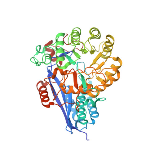
1JDF

Glucarate Dehydratase from E.coli N341D mutant


Experimental Data Snapshot

  • Method: X-RAY DIFFRACTION
  • Resolution: 2.00 Å
  • R-Value Free: 0.233 
  • R-Value Work: 0.198 
  • R-Value Observed: 0.198 

Starting Model: experimental
View more details

wwPDB Validation   3D Report Full Report


Ligand Structure Quality Assessment 


This is version 1.4 of the entry. See complete history


Literature

Evolution of enzymatic activities in the enolase superfamily: identification of the general acid catalyst in the active site of D-glucarate dehydratase from Escherichia coli.

Gulick, A.M.Hubbard, B.K.Gerlt, J.A.Rayment, I.

(2001) Biochemistry 40: 10054-10062

  • DOI: https://doi.org/10.1021/bi010733b
  • Primary Citation of Related Structures:  
    1JCT, 1JDF

  • PubMed Abstract: 

    D-Glucarate dehydratase from Escherichia coli (GlucD), a member of the enolase superfamily, catalyzes the dehydration of both D-glucarate and L-idarate to form 5-keto-4-deoxy-D-glucarate (KDG). Previous mutagenesis and structural studies identified Lys 207 and the His 339-Asp 313 dyad as the general basic catalysts that abstract the C5 proton from L-idarate and D-glucarate, respectively, thereby initiating the reaction by formation of a stabilized enediolate anion intermediate [Gulick, A. M., Hubbard, B. K., Gerlt, J. A., and Rayment, I. (2000) Biochemistry 39, 4590-4602]. The vinylogous elimination of the 4-OH group from this intermediate presumably requires a general acid catalyst. The structure of GlucD with KDG and 4-deoxy-D-glucarate bound in the active site revealed that only His 339 and Asn 341 are proximal to the presumed position of the 4-OH leaving group. The N341D and N341L mutants of GlucD were constructed and subjected to both mechanistic and structural analyses. The N341L but not N341D mutant catalyzed the dehydrofluorination of 4-deoxy-4-fluoro-D-glucarate, demonstrating that in this mutant the initial proton abstraction from C5 can be decoupled from elimination of the leaving group from C4. The kinetic properties and structures of these mutants suggest that either Asn 341 participates in catalysis as the general acid that facilitates the departure of the 4-leaving group or is essential for proper positioning of His 339. In the latter scenario, His 339 would function not only as the general base that abstracts the C5 proton from D-glucarate but also as the general acid that catalyzes both the departure of the 4-OH group and the stereospecific incorporation of solvent hydrogen with retention of configuration to form the KDG product. The involvement of a single functional group in this reaction highlights the plasticity of the active site design in members of the enolase superfamily.


  • Organizational Affiliation

    Department of Biochemistry, University of Wisconsin, Madison, Wisconsin 53706, USA.


Macromolecules
Find similar proteins by:  (by identity cutoff)  |  3D Structure
Entity ID: 1
MoleculeChains Sequence LengthOrganismDetailsImage
Glucarate Dehydratase
A, B, C, D
446Escherichia coliMutation(s): 1 
EC: 4.2.1.40
UniProt
Find proteins for P0AES2 (Escherichia coli (strain K12))
Explore P0AES2 
Go to UniProtKB:  P0AES2
Entity Groups  
Sequence Clusters30% Identity50% Identity70% Identity90% Identity95% Identity100% Identity
UniProt GroupP0AES2
Sequence Annotations
Expand
  • Reference Sequence
Small Molecules
Ligands 3 Unique
IDChains Name / Formula / InChI Key2D Diagram3D Interactions
GLR
Query on GLR

Download Ideal Coordinates CCD File 
F [auth A],
J [auth B],
N [auth C],
R [auth D]
2,3-DIHYDROXY-5-OXO-HEXANEDIOATE
C6 H6 O7
QUURPCHWPQNNGL-ZAFYKAAXSA-L
IPA
Query on IPA

Download Ideal Coordinates CCD File 
G [auth A]
H [auth A]
K [auth B]
L [auth B]
O [auth C]
G [auth A],
H [auth A],
K [auth B],
L [auth B],
O [auth C],
P [auth C],
S [auth D],
T [auth D]
ISOPROPYL ALCOHOL
C3 H8 O
KFZMGEQAYNKOFK-UHFFFAOYSA-N
MG
Query on MG

Download Ideal Coordinates CCD File 
E [auth A],
I [auth B],
M [auth C],
Q [auth D]
MAGNESIUM ION
Mg
JLVVSXFLKOJNIY-UHFFFAOYSA-N
Experimental Data & Validation

Experimental Data

  • Method: X-RAY DIFFRACTION
  • Resolution: 2.00 Å
  • R-Value Free: 0.233 
  • R-Value Work: 0.198 
  • R-Value Observed: 0.198 
  • Space Group: P 1
Unit Cell:
Length ( Å )Angle ( ˚ )
a = 70.925α = 104.11
b = 83.926β = 93.75
c = 98.222γ = 113.12
Software Package:
Software NamePurpose
DENZOdata reduction
SCALEPACKdata scaling
CNSrefinement
CNSphasing

Structure Validation

View Full Validation Report


Ligand Structure Quality Assessment 


Entry History 

Deposition Data

Revision History  (Full details and data files)

  • Version 1.0: 2001-09-05
    Type: Initial release
  • Version 1.1: 2008-04-27
    Changes: Version format compliance
  • Version 1.2: 2011-07-13
    Changes: Version format compliance
  • Version 1.3: 2021-10-27
    Changes: Database references, Derived calculations
  • Version 1.4: 2023-08-16
    Changes: Data collection, Refinement description