1JB8 | pdb_00001jb8

The Crystal Structure of an RNA/DNA Hybrid Reveals Novel Intermolecular Intercalation


Experimental Data Snapshot

  • Method: X-RAY DIFFRACTION
  • Resolution: 2.38 Å
  • R-Value Free: 
    0.251 (Depositor) 
  • R-Value Work: 
    0.223 (Depositor), 0.238 (DCC) 

Starting Model: experimental
View more details

wwPDB Validation 3D Report Full Report

Validation slider image for 1JB8

This is version 1.5 of the entry. See complete history

Literature

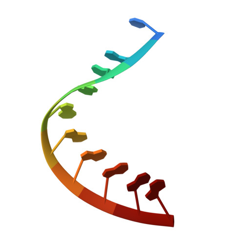
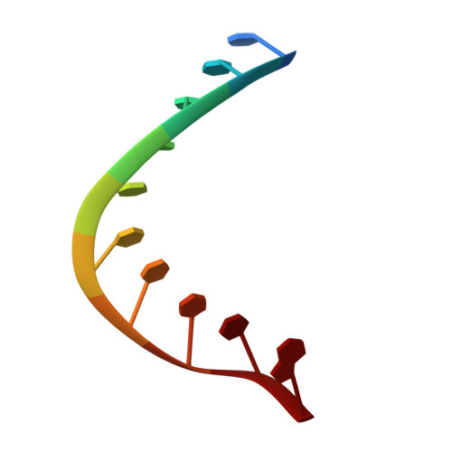
Crystal structure of an RNADNA hybrid reveals intermolecular intercalation: Dimer formation by base-pair swapping

Han, G.W.Kopka, M.L.Langs, D.Sawaya, M.R.Dickerson, R.E.

(2003) Proc Natl Acad Sci U S A 100: 9214-9219

  • DOI: https://doi.org/10.1073/pnas.1533326100
  • Primary Citation Related Structures: 
    1JB8

  • PubMed Abstract: 

    An intermolecular intercalation of base pairs was found at the CA step in the I222 crystal structure of the RNA.DNA hybrid, r(CAAAGAAAAG).d(CTTTTCTTTG), which contains two-thirds of the polypurine tract sequence of HIV-1 with a substitution of cytosine for the initial adenine. This sequence crystallized in both P212121 and I222 space groups, with an rms difference of only 0.63 A between residues 3 to 18 of the two forms. P212121 and I222 helices are both A-like, but intercalation occurs only in the I222 crystal form. The present structure shows bases stacked in parallel rather than perpendicular as in intercalated DNA (I-DNA). The base intercalation is also different from zipper-like meshing of bases seen in the center of the crystal structure of d(GCGAAAGCT), which does not have Watson-Crick base pairing. The base-step intercalation seen here is reminiscent of domain swapping in proteins; therefore, we call this phenomenon "base-pair swapping." It involves a highly mobile CA step and seems to be sequence-specific and electrostatically stable without disrupting Watson-Crick interactions. It also exhibits a large rise concurrent with unwinding of the helix (low twist). We present a base-pair swapping dimer in nucleic acids.


  • Organizational Affiliation
    • Molecular Biology Institute, University of California, Los Angeles, CA 90095-1570, USA.

Macromolecule Content 

  • Total Structure Weight: 6.25 kDa 
  • Atom Count: 480 
  • Modeled Residue Count: 20 
  • Deposited Residue Count: 20 
  • Unique nucleic acid chains: 2

Macromolecules

Find similar nucleic acids by:  Sequence
Entity ID: 1
MoleculeChains LengthOrganismImage
5'-R(*CP*AP*AP*AP*GP*AP*AP*AP*AP*G)-3'10N/A
Sequence Annotations
Expand
Reference Sequence
Find similar nucleic acids by:  Sequence
Entity ID: 2
MoleculeChains LengthOrganismImage
5'-D(*CP*TP*TP*TP*TP*CP*TP*TP*TP*G)-3'10N/A
Sequence Annotations
Expand
Reference Sequence

Experimental Data & Validation

Experimental Data

  • Method: X-RAY DIFFRACTION
  • Resolution: 2.38 Å
  • R-Value Free:  0.251 (Depositor) 
  • R-Value Work:  0.223 (Depositor), 0.238 (DCC) 
Space Group: I 2 2 2
Unit Cell:
Length ( Å )Angle ( ˚ )
a = 45.234α = 90
b = 46.78β = 90
c = 56.56γ = 90
Software Package:
Software NamePurpose
DENZOdata reduction
EPMRphasing
CNSrefinement

Structure Validation

View Full Validation Report


Entry History 

Deposition Data

Revision History  (Full details and data files)

  • Version 1.0: 2003-07-01
    Type: Initial release
  • Version 1.1: 2008-04-27
    Changes: Version format compliance
  • Version 1.2: 2011-07-13
    Changes: Version format compliance
  • Version 1.3: 2017-10-04
    Changes: Refinement description
  • Version 1.4: 2024-02-07
    Changes: Data collection, Database references
  • Version 1.5: 2024-04-03
    Changes: Refinement description