1J6Z | pdb_00001j6z

UNCOMPLEXED ACTIN


Experimental Data Snapshot

  • Method: X-RAY DIFFRACTION
  • Resolution: 1.54 Å
  • R-Value Free: 
    0.223 (Depositor) 
  • R-Value Work: 
    0.179 (Depositor) 
  • R-Value Observed: 
    0.179 (Depositor) 

Starting Model: experimental
View more details

wwPDB Validation 3D Report Full Report

Validation slider image for 1J6Z

This is version 1.4 of the entry. See complete history

Literature

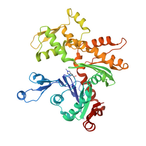
The crystal structure of uncomplexed actin in the ADP state.

Otterbein, L.R.Graceffa, P.Dominguez, R.

(2001) Science 293: 708-711

  • DOI: https://doi.org/10.1126/science.1059700
  • Primary Citation Related Structures: 
    1J6Z

  • PubMed Abstract: 

    The dynamics and polarity of actin filaments are controlled by a conformational change coupled to the hydrolysis of adenosine 5'-triphosphate (ATP) by a mechanism that remains to be elucidated. Actin modified to block polymerization was crystallized in the adenosine 5'-diphosphate (ADP) state, and the structure was solved to 1.54 angstrom resolution. Compared with previous ATP-actin structures from complexes with deoxyribonuclease I, profilin, and gelsolin, monomeric ADP-actin is characterized by a marked conformational change in subdomain 2. The successful crystallization of monomeric actin opens the way to future structure determinations of actin complexes with actin-binding proteins such as myosin.


  • Organizational Affiliation
    • Boston Biomedical Research Institute, 64 Grove Street, Watertown, MA 02472, USA.

Macromolecule Content 

  • Total Structure Weight: 43.03 kDa 
  • Atom Count: 3,350 
  • Modeled Residue Count: 369 
  • Deposited Residue Count: 375 
  • Unique protein chains: 1

Macromolecules

Find similar proteins by:|  3D Structure
Entity ID: 1
MoleculeChains  Sequence LengthOrganismDetailsImage
ACTIN ALPHA 1375Oryctolagus cuniculusMutation(s): 1 
EC: 3.6.4
UniProt
Find proteins for P68135 (Oryctolagus cuniculus)
Explore P68135 
Go to UniProtKB:  P68135
Entity Groups
Sequence Clusters30% Identity50% Identity70% Identity90% Identity95% Identity100% Identity
UniProt GroupP68135
Sequence Annotations
Expand
Reference Sequence

Small Molecules

Ligands 3 Unique
IDChains Name / Formula / InChI Key2D Diagram3D Interactions
RHO

Query on RHO


Download:Ideal Coordinates CCD File
I [auth A]TETRAMETHYLRHODAMINE-5-MALEIMIDE
C28 H25 N3 O5
KGFLZYXDJDOIEE-UHFFFAOYSA-N
ADP

Query on ADP


Download:Ideal Coordinates CCD File
H [auth A]ADENOSINE-5'-DIPHOSPHATE
C10 H15 N5 O10 P2
XTWYTFMLZFPYCI-KQYNXXCUSA-N
CA

Query on CA


Download:Ideal Coordinates CCD File
B [auth A]
C [auth A]
D [auth A]
E [auth A]
F [auth A]
B [auth A],
C [auth A],
D [auth A],
E [auth A],
F [auth A],
G [auth A]
CALCIUM ION
Ca
BHPQYMZQTOCNFJ-UHFFFAOYSA-N
Modified Residues  1 Unique
IDChains TypeFormula2D DiagramParent
HIC
Query on HIC
A
L-PEPTIDE LINKINGC7 H11 N3 O2HIS

Experimental Data & Validation

Experimental Data

  • Method: X-RAY DIFFRACTION
  • Resolution: 1.54 Å
  • R-Value Free:  0.223 (Depositor) 
  • R-Value Work:  0.179 (Depositor) 
  • R-Value Observed: 0.179 (Depositor) 
Space Group: C 1 2 1
Unit Cell:
Length ( Å )Angle ( ˚ )
a = 112.808α = 90
b = 37.498β = 108.26
c = 85.261γ = 90
Software Package:
Software NamePurpose
HKL-2000data collection
HKL-2000data reduction
AMoREphasing
WARPmodel building
REFMACrefinement
HKL-2000data scaling
ARP/wARPmodel building

Structure Validation

View Full Validation Report


Entry History 

Deposition Data

Revision History  (Full details and data files)

  • Version 1.0: 2001-08-15
    Type: Initial release
  • Version 1.1: 2008-04-27
    Changes: Version format compliance
  • Version 1.2: 2011-07-13
    Changes: Version format compliance
  • Version 1.3: 2017-10-04
    Changes: Refinement description
  • Version 1.4: 2023-08-16
    Changes: Data collection, Database references, Derived calculations, Refinement description