1J53 | pdb_00001j53

Structure of the N-terminal Exonuclease Domain of the Epsilon Subunit of E.coli DNA Polymerase III at pH 8.5


Experimental Data Snapshot

  • Method: X-RAY DIFFRACTION
  • Resolution: 1.80 Å
  • R-Value Free: 
    0.227 (Depositor), 0.251 (DCC) 
  • R-Value Work: 
    0.199 (Depositor), 0.216 (DCC) 

wwPDB Validation 3D Report Full Report

Validation slider image for 1J53

Ligand Structure Quality Assessment 


This is version 1.4 of the entry. See complete history

Literature

Structural Basis for Proofreading during Replication of the Escherichia coli Chromosome

Hamdan, S.Carr, P.D.Brown, S.E.Ollis, D.L.Dixon, N.E.

(2002) Structure 10: 535-546

  • DOI: https://doi.org/10.1016/s0969-2126(02)00738-4
  • Primary Citation Related Structures: 
    1J53, 1J54

  • PubMed Abstract: 

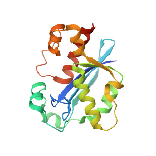
    The epsilon subunit of the Escherichia coli replicative DNA polymerase III is the proofreading 3'-5' exonuclease. Structures of its catalytic N-terminal domain (epsilon186) were determined at two pH values (5.8 and 8.5) at resolutions of 1.7-1.8 A, in complex with two Mn(II) ions and a nucleotide product of its reaction, thymidine 5'-monophosphate. The protein structure is built around a core five-stranded beta sheet that is a common feature of members of the DnaQ superfamily. The structures were identical, except for differences in the way TMP and water molecules are coordinated to the binuclear metal center in the active site. These data are used to develop a mechanism for epsilon and to produce a plausible model of the complex of epsilon186 with DNA.


  • Organizational Affiliation
    • Research School of Chemistry, Australian National University, Canberra, ACT, Australia.

Macromolecule Content 

  • Total Structure Weight: 21.62 kDa 
  • Atom Count: 1,641 
  • Modeled Residue Count: 174 
  • Deposited Residue Count: 186 
  • Unique protein chains: 1

Macromolecules

Find similar proteins by:|  3D Structure
Entity ID: 1
MoleculeChains  Sequence LengthOrganismDetailsImage
DNA polymerase III, epsilon chain186Escherichia coliMutation(s): 0 
EC: 2.7.7.7
UniProt
Find proteins for P03007 (Escherichia coli (strain K12))
Explore P03007 
Go to UniProtKB:  P03007
Entity Groups
Sequence Clusters30% Identity50% Identity70% Identity90% Identity95% Identity100% Identity
UniProt GroupP03007
Sequence Annotations
Expand
Reference Sequence

Experimental Data & Validation

Experimental Data

  • Method: X-RAY DIFFRACTION
  • Resolution: 1.80 Å
  • R-Value Free:  0.227 (Depositor), 0.251 (DCC) 
  • R-Value Work:  0.199 (Depositor), 0.216 (DCC) 
Space Group: P 41 21 2
Unit Cell:
Length ( Å )Angle ( ˚ )
a = 60.8α = 90
b = 60.8β = 90
c = 111.1γ = 90
Software Package:
Software NamePurpose
DENZOdata reduction
SCALEPACKdata scaling
CCP4model building
CNSrefinement
CCP4phasing

Structure Validation

View Full Validation Report


Ligand Structure Quality Assessment 


Entry History 

Deposition Data

Revision History  (Full details and data files)

  • Version 1.0: 2002-10-16
    Type: Initial release
  • Version 1.1: 2008-04-26
    Changes: Version format compliance
  • Version 1.2: 2011-07-13
    Changes: Version format compliance
  • Version 1.3: 2023-12-27
    Changes: Data collection, Database references, Derived calculations
  • Version 1.4: 2024-12-25
    Changes: Advisory, Derived calculations, Structure summary