1J3L | pdb_00001j3l

Structure of the RNA-processing inhibitor RraA from Thermus thermophilis


Experimental Data Snapshot

  • Method: X-RAY DIFFRACTION
  • Resolution: 2.30 Å
  • R-Value Free: 
    0.274 (Depositor), 0.273 (DCC) 
  • R-Value Work: 
    0.206 (Depositor), 0.202 (DCC) 
  • R-Value Observed: 
    0.210 (Depositor) 

wwPDB Validation 3D Report Full Report

Validation slider image for 1J3L

This is version 1.4 of the entry. See complete history

Literature

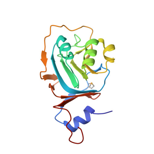
Structure of the RNA-processing inhibitor RraA from Thermus thermophilis.

Rehse, P.H.Kuroishi, C.Tahirov, T.H.

(2004) Acta Crystallogr D Biol Crystallogr 60: 1997-2002

  • DOI: https://doi.org/10.1107/S0907444904021146
  • Primary Citation Related Structures: 
    1J3L

  • PubMed Abstract: 

    The menG gene product, thought to catalyze the final methylation in vitamin K(2) synthesis, has recently been shown to inhibit RNase E in Eschericha coli. The structure of the protein, since renamed RraA, has been solved to 2.3 A using the multiple-wavelength anomalous diffraction method and selenomethionine-substituted protein from Thermus thermophilus. The six molecules in the asymmetric unit are arranged as two similar trimers which have a degree of interaction, suggesting biological significance. The fold does not support the postulated methylation function. Genomic analysis, specifically a lack of an RNase E homologue in cases where homologues to RraA exist, indicates that the function is still obscure.


  • Organizational Affiliation
    • Highthroughput Factory, RIKEN Harima Institute, 1-1-1 Kouto, Mikazuki-cho, Sayo-gun, Hyogo 679-5148, Japan.

Macromolecule Content 

  • Total Structure Weight: 104.84 kDa 
  • Atom Count: 7,692 
  • Modeled Residue Count: 974 
  • Deposited Residue Count: 984 
  • Unique protein chains: 1

Macromolecules

Find similar proteins by:|  3D Structure
Entity ID: 1
MoleculeChains  Sequence LengthOrganismDetailsImage
Demethylmenaquinone Methyltransferase
A, B, C, D, E
A, B, C, D, E, F
164Thermus thermophilusMutation(s): 0 
EC: 2.1.1 (PDB Primary Data), 4.1.3.17 (UniProt), 4.1.1.112 (UniProt)
UniProt
Find proteins for Q5SIP7 (Thermus thermophilus (strain ATCC 27634 / DSM 579 / HB8))
Explore Q5SIP7 
Go to UniProtKB:  Q5SIP7
Entity Groups
Sequence Clusters30% Identity50% Identity70% Identity90% Identity95% Identity100% Identity
UniProt GroupQ5SIP7
Sequence Annotations
Expand
Reference Sequence

Small Molecules

Modified Residues  1 Unique
IDChains TypeFormula2D DiagramParent
MSE
Query on MSE
A, B, C, D, E
A, B, C, D, E, F
L-PEPTIDE LINKINGC5 H11 N O2 SeMET

Experimental Data & Validation

Experimental Data

  • Method: X-RAY DIFFRACTION
  • Resolution: 2.30 Å
  • R-Value Free:  0.274 (Depositor), 0.273 (DCC) 
  • R-Value Work:  0.206 (Depositor), 0.202 (DCC) 
  • R-Value Observed: 0.210 (Depositor) 
Space Group: C 2 2 21
Unit Cell:
Length ( Å )Angle ( ˚ )
a = 61.872α = 90
b = 109.068β = 90
c = 270.319γ = 90
Software Package:
Software NamePurpose
CNSrefinement
DENZOdata reduction
SCALEPACKdata scaling
SOLVEphasing

Structure Validation

View Full Validation Report


Entry History 

Deposition Data

Revision History  (Full details and data files)

  • Version 1.0: 2004-02-17
    Type: Initial release
  • Version 1.1: 2008-04-27
    Changes: Version format compliance
  • Version 1.2: 2011-07-13
    Changes: Version format compliance
  • Version 1.3: 2023-12-27
    Changes: Data collection, Database references, Derived calculations
  • Version 1.4: 2024-10-30
    Changes: Structure summary