1J1O | pdb_00001j1o

Crystal Structure of HyHEL-10 Fv mutant LY50F complexed with hen egg white lysozyme


Experimental Data Snapshot

  • Method: X-RAY DIFFRACTION
  • Resolution: 1.80 Å
  • R-Value Free: 
    0.215 (Depositor), 0.190 (DCC) 
  • R-Value Work: 
    0.190 (Depositor), 0.190 (DCC) 

wwPDB Validation   3D Report Full Report


This is version 1.5 of the entry. See complete history


Literature

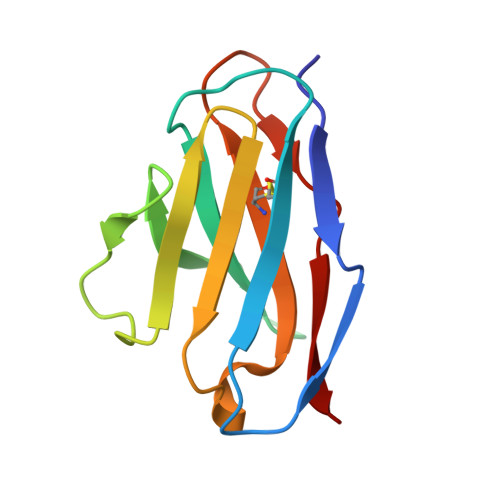
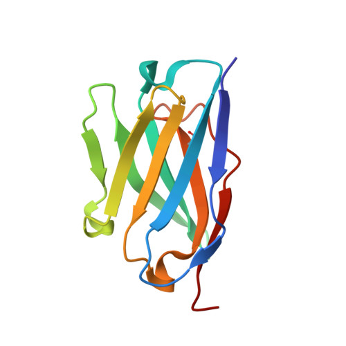
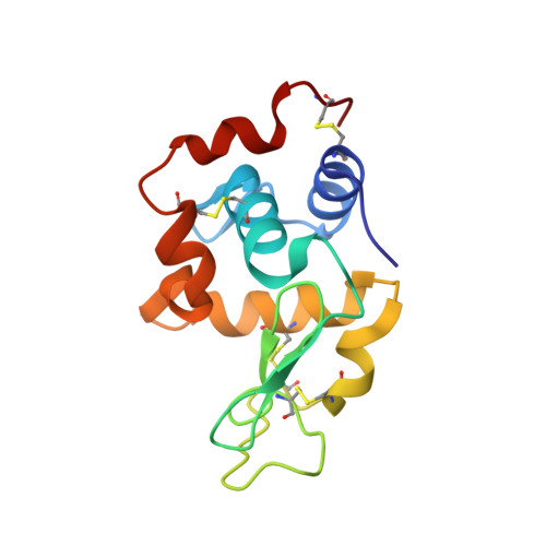
The Role of Hydrogen Bonding via Interfacial Water Molecules in Antigen-Antibody Complexation. THE HyHEL-10-HEL INTERACTION

Yokota, A.Tsumoto, K.Shiroishi, M.Kondo, H.Kumagai, I.

(2003) J Biological Chem 278: 5410-5418

  • DOI: https://doi.org/10.1074/jbc.M210182200
  • Primary Citation of Related Structures:  
    1J1O, 1J1P, 1J1X

  • PubMed Abstract: 

    To study the role of hydrogen bonding via interfacial water molecules in protein-protein interactions, we examined the interaction between hen egg white lysozyme (HEL) and its HyHEL-10 variable domain fragment (Fv) antibody. We constructed three antibody mutants (l-Y50F, l-S91A, and l-S93A) and investigated the interactions between the mutant Fvs and HEL. Isothermal titration calorimetry indicated that the mutations significantly decreased the negative enthalpy change (8-25 kJ mol(-1)), despite some offset by a favorable entropy change. X-ray crystallography demonstrated that the complexes had nearly identical structures, including the positions of the interfacial water molecules. Taken together, the isothermal titration calorimetric and x-ray crystallographic results indicate that hydrogen bonding via interfacial water enthalpically contributes to the Fv-HEL interaction despite the partial offset because of entropy loss, suggesting that hydrogen bonding stiffens the antigen-antibody complex.


  • Organizational Affiliation

    Department of Biomolecular Engineering, Graduate School of Engineering, Tohoku University, Aoba-yama 07, Aoba-ku, Sendai 980-8579, Japan.


Macromolecules
Find similar proteins by:  (by identity cutoff)  |  3D Structure
Entity ID: 1
MoleculeChains Sequence LengthOrganismDetailsImage
lysozyme binding Ig kappa chain V23-J2 regionA [auth L]107Mus musculusMutation(s): 1 
UniProt
Find proteins for P01642 (Mus musculus)
Explore P01642 
Go to UniProtKB:  P01642
Entity Groups  
Sequence Clusters30% Identity50% Identity70% Identity90% Identity95% Identity100% Identity
UniProt GroupP01642
Sequence Annotations
Expand
  • Reference Sequence
Find similar proteins by:  (by identity cutoff)  |  3D Structure
Entity ID: 2
MoleculeChains Sequence LengthOrganismDetailsImage
Ig VH,anti-lysozymeB [auth H]114Mus musculusMutation(s): 0 
UniProt
Find proteins for P01823 (Mus musculus)
Explore P01823 
Go to UniProtKB:  P01823
Entity Groups  
Sequence Clusters30% Identity50% Identity70% Identity90% Identity95% Identity100% Identity
UniProt GroupP01823
Sequence Annotations
Expand
  • Reference Sequence
Find similar proteins by:  (by identity cutoff)  |  3D Structure
Entity ID: 3
MoleculeChains Sequence LengthOrganismDetailsImage
Lysozyme CC [auth Y]129Gallus gallusMutation(s): 0 
EC: 3.2.1.17
UniProt
Find proteins for P00698 (Gallus gallus)
Explore P00698 
Go to UniProtKB:  P00698
Entity Groups  
Sequence Clusters30% Identity50% Identity70% Identity90% Identity95% Identity100% Identity
UniProt GroupP00698
Sequence Annotations
Expand
  • Reference Sequence
Experimental Data & Validation

Experimental Data

  • Method: X-RAY DIFFRACTION
  • Resolution: 1.80 Å
  • R-Value Free:  0.215 (Depositor), 0.190 (DCC) 
  • R-Value Work:  0.190 (Depositor), 0.190 (DCC) 
Space Group: P 41 21 2
Unit Cell:
Length ( Å )Angle ( ˚ )
a = 56.23α = 90
b = 56.23β = 90
c = 234.486γ = 90
Software Package:
Software NamePurpose
MOSFLMdata reduction
SCALAdata scaling
CNSrefinement
CCP4data scaling
CNSphasing

Structure Validation

View Full Validation Report


Entry History 

Deposition Data

Revision History  (Full details and data files)

  • Version 1.0: 2003-01-14
    Type: Initial release
  • Version 1.1: 2008-04-27
    Changes: Version format compliance
  • Version 1.2: 2011-07-13
    Changes: Version format compliance
  • Version 1.3: 2021-11-10
    Changes: Database references
  • Version 1.4: 2023-12-27
    Changes: Data collection
  • Version 1.5: 2024-11-06
    Changes: Structure summary