1IY8 | pdb_00001iy8

Crystal Structure of Levodione Reductase


Experimental Data Snapshot

  • Method: X-RAY DIFFRACTION
  • Resolution: 1.60 Å
  • R-Value Free: 
    0.219 (Depositor), 0.219 (DCC) 
  • R-Value Work: 
    0.199 (Depositor), 0.199 (DCC) 

Starting Model: experimental
View more details

wwPDB Validation 3D Report Full Report

Validation slider image for 1IY8

Ligand Structure Quality Assessment 


This is version 1.3 of the entry. See complete history

Literature

The Crystal Structure and Stereospecificity of Levodione Reductase from Corynebacterium aquaticum M-13

Sogabe, S.Yoshizumi, A.Fukami, T.Shiratori, Y.Shimizu, S.Takagi, H.Nakamori, S.Wada, M.

(2003) J Biological Chem 278: 19387-19395

  • DOI: https://doi.org/10.1074/jbc.M208146200
  • Primary Citation Related Structures: 
    1IY8

  • PubMed Abstract: 

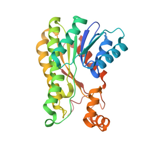
    The (6R)-2,2,6-trimethyl-1,4-cyclohexanedione (levodione) reductase (LVR) of the soil isolate bacterium Corynebacterium aquaticum M-13 is a NAD(H)-linked enzyme that catalyzes reversible oxidoreduction between (4R)-hydroxy-(6R)-2,2,6-trimethylcyclohexanone (actinol) and levodione. Here the crystal structure of a ternary complex of LVR with NADH and its inhibitor 2-methyl-2,4-pentanediol has been determined by molecular replacement and refined at 1.6-A resolution with a crystallographic R factor of 0.199. The overall structure is similar to those of other short-chain alcohol dehydrogenase/reductase enzymes. The positions of NADH and 2-methyl-2,4-pentanediol indicate the binding site of the substrate and identify residues that are likely to be important in the catalytic reaction. Modeling of the substrate binding in the active site suggests that the specificity of LVR is determined by electrostatic interactions between the negatively charged surface of Glu-103 of LVR and the positively charged surface on the re side of levodione. Mutant LVR enzymes in which Glu-103 is substituted with alanine (E103A), glutamine (E103Q), asparagines (E103N), or aspartic acid (E103D) show a 2-6-fold increase in Km values as compared with wild-type LVR and a much lower enantiomeric excess of the reaction products (60%) than the wild-type enzyme (95%). Together, these data indicate that Glu-103 has an important role in determining the stereospecificity of LVR.


  • Organizational Affiliation
    • Nippon Roche Research Center, 200 Kajiwara, Kamakura, Kanagawa 247-8530, Japan.

Macromolecule Content 

  • Total Structure Weight: 229.82 kDa 
  • Atom Count: 16,602 
  • Modeled Residue Count: 2,064 
  • Deposited Residue Count: 2,136 
  • Unique protein chains: 1

Macromolecules

Find similar proteins by:|  3D Structure
Entity ID: 1
MoleculeChains  Sequence LengthOrganismDetailsImage
LEVODIONE REDUCTASE
A, B, C, D, E
A, B, C, D, E, F, G, H
267Leifsonia aquaticaMutation(s): 0 
EC: 1.1.1
UniProt
Find proteins for Q9LBG2 (Leifsonia aquatica)
Explore Q9LBG2 
Go to UniProtKB:  Q9LBG2
Entity Groups
Sequence Clusters30% Identity50% Identity70% Identity90% Identity95% Identity100% Identity
UniProt GroupQ9LBG2
Sequence Annotations
Expand
Reference Sequence

Small Molecules

Ligands 2 Unique
IDChains Name / Formula / InChI Key2D Diagram3D Interactions
NAD

Query on NAD


Download:Ideal Coordinates CCD File
I [auth A]
K [auth B]
M [auth C]
O [auth D]
Q [auth E]
I [auth A],
K [auth B],
M [auth C],
O [auth D],
Q [auth E],
S [auth F],
U [auth G],
W [auth H]
NICOTINAMIDE-ADENINE-DINUCLEOTIDE
C21 H27 N7 O14 P2
BAWFJGJZGIEFAR-NNYOXOHSSA-N
MRD

Query on MRD


Download:Ideal Coordinates CCD File
J [auth A]
L [auth B]
N [auth C]
P [auth D]
R [auth E]
J [auth A],
L [auth B],
N [auth C],
P [auth D],
R [auth E],
T [auth F],
V [auth G],
X [auth H]
(4R)-2-METHYLPENTANE-2,4-DIOL
C6 H14 O2
SVTBMSDMJJWYQN-RXMQYKEDSA-N

Experimental Data & Validation

Experimental Data

  • Method: X-RAY DIFFRACTION
  • Resolution: 1.60 Å
  • R-Value Free:  0.219 (Depositor), 0.219 (DCC) 
  • R-Value Work:  0.199 (Depositor), 0.199 (DCC) 
Space Group: P 1
Unit Cell:
Length ( Å )Angle ( ˚ )
a = 66.672α = 103.87
b = 79.184β = 91.4
c = 112.67γ = 105.48
Software Package:
Software NamePurpose
MOSFLMdata reduction
SCALAdata scaling
CNSrefinement
CCP4data scaling
CNSphasing

Structure Validation

View Full Validation Report


Ligand Structure Quality Assessment 


Entry History 

Deposition Data

Revision History  (Full details and data files)

  • Version 1.0: 2003-05-27
    Type: Initial release
  • Version 1.1: 2008-04-27
    Changes: Version format compliance
  • Version 1.2: 2011-07-13
    Changes: Version format compliance
  • Version 1.3: 2023-10-25
    Changes: Data collection, Database references, Derived calculations, Refinement description