1IWO | pdb_00001iwo

Crystal structure of the SR Ca2+-ATPase in the absence of Ca2+


Experimental Data Snapshot

  • Method: X-RAY DIFFRACTION
  • Resolution: 3.10 Å
  • R-Value Free: 
    0.268 (Depositor) 
  • R-Value Work: 
    0.237 (Depositor) 
  • R-Value Observed: 
    0.237 (Depositor) 

Starting Model: experimental
View more details

wwPDB Validation 3D Report Full Report

Validation slider image for 1IWO

This is version 1.4 of the entry. See complete history

Literature

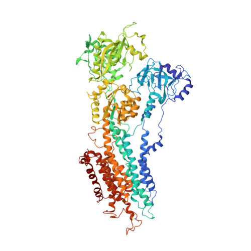
Structural changes in the calcium pump accompanying the dissociation of calcium

Toyoshima, C.Nomura, H.

(2002) Nature 418: 605-611

  • DOI: https://doi.org/10.1038/nature00944
  • Primary Citation Related Structures: 
    1IWO

  • PubMed Abstract: 

    In skeletal muscle, calcium ions are transported (pumped) against a concentration gradient from the cytoplasm into the sarcoplasmic reticulum, an intracellular organelle. This causes muscle cells to relax after cytosolic calcium increases during excitation. The Ca(2+) ATPase that carries out this pumping is a representative P-type ion-transporting ATPase. Here we describe the structure of this ion pump at 3.1 A resolution in a Ca(2+)-free (E2) state, and compare it with that determined previously for the Ca(2+)-bound (E1Ca(2+)) state. The structure of the enzyme stabilized by thapsigargin, a potent inhibitor, shows large conformation differences from that in E1Ca(2+). Three cytoplasmic domains gather to form a single headpiece, and six of the ten transmembrane helices exhibit large-scale rearrangements. These rearrangements ensure the release of calcium ions into the lumen of sarcoplasmic reticulum and, on the cytoplasmic side, create a pathway for entry of new calcium ions.


  • Organizational Affiliation
    • Institute of Molecular and Cellular Biosciences, The University of Tokyo, Bunkyo-ku, Tokyo 113-0032, Japan. ct@iam.u-tokyo.ac.jp

Macromolecule Content 

  • Total Structure Weight: 220.51 kDa 
  • Atom Count: 15,434 
  • Modeled Residue Count: 1,988 
  • Deposited Residue Count: 1,988 
  • Unique protein chains: 1

Macromolecules

Find similar proteins by:|  3D Structure
Entity ID: 1
MoleculeChains  Sequence LengthOrganismDetailsImage
Sarcoplasmic/endoplasmic reticulum calcium ATPase 1
A, B
994Oryctolagus cuniculusMutation(s): 0 
EC: 7.2.2.10
Membrane Entity: Yes 
UniProt
Find proteins for P04191 (Oryctolagus cuniculus)
Explore P04191 
Go to UniProtKB:  P04191
Entity Groups
Sequence Clusters30% Identity50% Identity70% Identity90% Identity95% Identity100% Identity
UniProt GroupP04191
Sequence Annotations
Expand
Reference Sequence

Small Molecules

Ligands 1 Unique
IDChains Name / Formula / InChI Key2D Diagram3D Interactions
TG1

Query on TG1


Download:Ideal Coordinates CCD File
C [auth A],
D [auth B]
OCTANOIC ACID [3S-[3ALPHA, 3ABETA, 4ALPHA, 6BETA, 6ABETA, 7BETA, 8ALPHA(Z), 9BALPHA]]-6-(ACETYLOXY)-2,3,-3A,4,5,6,6A,7,8,9B-DECAHYDRO-3,3A-DIHYDROXY-3,6,9-TRIMETHYL-8-[(2-METHYL-1-OXO-2-BUTENYL)OX Y]-2-OXO-4-(1-OXOBUTOXY)-AZULENO[4,5-B]FURAN-7-YL ESTER
C34 H50 O12
IXFPJGBNCFXKPI-FSIHEZPISA-N

Experimental Data & Validation

Experimental Data

  • Method: X-RAY DIFFRACTION
  • Resolution: 3.10 Å
  • R-Value Free:  0.268 (Depositor) 
  • R-Value Work:  0.237 (Depositor) 
  • R-Value Observed: 0.237 (Depositor) 
Space Group: P 41
Unit Cell:
Length ( Å )Angle ( ˚ )
a = 71.739α = 90
b = 71.739β = 90
c = 590.3γ = 90
Software Package:
Software NamePurpose
DENZOdata reduction
SCALEPACKdata scaling
X-PLORmodel building
CNSrefinement
X-PLORphasing

Structure Validation

View Full Validation Report


Entry History 

Deposition Data

Revision History  (Full details and data files)

  • Version 1.0: 2002-08-14
    Type: Initial release
  • Version 1.1: 2008-04-27
    Changes: Version format compliance
  • Version 1.2: 2011-07-13
    Changes: Non-polymer description, Version format compliance
  • Version 1.3: 2023-10-25
    Changes: Data collection, Database references, Derived calculations, Refinement description, Structure summary
  • Version 1.4: 2024-10-23
    Changes: Structure summary