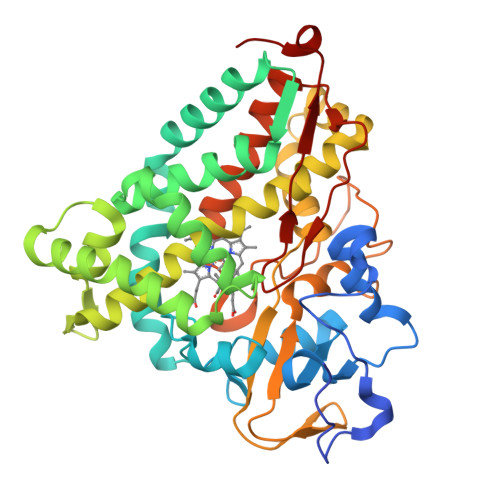
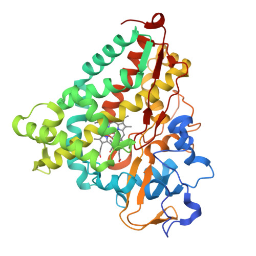
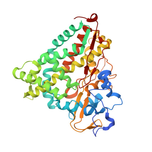
1IWI

Putidaredoxin-Binding Stablilizes an Active Conformer of Cytochrome P450cam in its Reduced State; Crystal Structure of Cytochrome P450cam


Experimental Data Snapshot

  • Method: X-RAY DIFFRACTION
  • Resolution: 2.00 Å
  • R-Value Free: 0.182 
  • R-Value Work: 0.177 

wwPDB Validation   3D Report Full Report


This is version 1.4 of the entry. See complete history


Literature

Infrared spectroscopic and mutational studies on putidaredoxin-induced conformational changes in ferrous CO-P450cam

Nagano, S.Shimada, H.Tarumi, A.Hishiki, T.Kimata-Ariga, Y.Egawa, T.Suematsu, M.Park, S.-Y.Adachi, S.Shiro, Y.Ishimura, Y.

(2003) Biochemistry 42: 14507-14514

  • DOI: https://doi.org/10.1021/bi035410p
  • Primary Citation of Related Structures:  
    1IWI, 1IWJ, 1IWK

  • PubMed Abstract: 

    Ferrous-carbon monoxide bound form of cytochrome P450cam (CO-P450cam) has two infrared (IR) CO stretching bands at 1940 and 1932 cm(-1). The former band is dominant (>95% in area) for CO-P450cam free of putidaredoxin (Pdx), while the latter band is dominant (>95% in area) in the complex of CO-P450cam with reduced Pdx. The binding of Pdx to CO-P450cam thus evokes a conformational change in the heme active site. To study the mechanism involved in the conformational change, surface amino acid residues Arg79, Arg109, and Arg112 in P450cam were replaced with Lys, Gln, and Met. IR spectroscopic and kinetic analyses of the mutants revealed that an enzyme that has a larger 1932 cm(-1) band area upon Pdx-binding has a larger catalytic activity. Examination of the crystal structures of R109K and R112K suggested that the interaction between the guanidium group of Arg112 and Pdx is important for the conformational change. The mutations did not change a coupling ratio between the hydroxylation product and oxygen consumed. We interpret these findings to mean that the interaction of P450cam with Pdx through Arg112 enhances electron donation from the proximal ligand (Cys357) to the O-O bond of iron-bound O(2) and, possibly, promotes electron transfer from reduced Pdx to oxyP450cam, thereby facilitating the O-O bond splitting.


  • Organizational Affiliation

    Department of Biochemistry and Integrative Medical Biology, School of Medicine, Keio University, 35 Shinano-machi, Shinjuku-ku, Tokyo 160-8582, Japan. snagano@uci.edu


Macromolecules
Find similar proteins by:  (by identity cutoff)  |  3D Structure
Entity ID: 1
MoleculeChains Sequence LengthOrganismDetailsImage
CYTOCHROME P450-CAM415Pseudomonas putidaMutation(s): 0 
EC: 1.14.15.1
UniProt
Find proteins for P00183 (Pseudomonas putida)
Explore P00183 
Go to UniProtKB:  P00183
Entity Groups  
Sequence Clusters30% Identity50% Identity70% Identity90% Identity95% Identity100% Identity
UniProt GroupP00183
Sequence Annotations
Expand
  • Reference Sequence
Small Molecules
Ligands 2 Unique
IDChains Name / Formula / InChI Key2D Diagram3D Interactions
HEM
Query on HEM

Download Ideal Coordinates CCD File 
B [auth A]PROTOPORPHYRIN IX CONTAINING FE
C34 H32 Fe N4 O4
KABFMIBPWCXCRK-RGGAHWMASA-L
CAM
Query on CAM

Download Ideal Coordinates CCD File 
C [auth A]CAMPHOR
C10 H16 O
DSSYKIVIOFKYAU-XCBNKYQSSA-N
Experimental Data & Validation

Experimental Data

  • Method: X-RAY DIFFRACTION
  • Resolution: 2.00 Å
  • R-Value Free: 0.182 
  • R-Value Work: 0.177 
  • Space Group: P 21 21 21
Unit Cell:
Length ( Å )Angle ( ˚ )
a = 109.16α = 90
b = 104.33β = 90
c = 36.18γ = 90
Software Package:
Software NamePurpose
MOSFLMdata reduction
SCALAdata scaling
X-PLORmodel building
X-PLORrefinement
CCP4data scaling
X-PLORphasing

Structure Validation

View Full Validation Report


Entry History 

Revision History  (Full details and data files)

  • Version 1.0: 2002-06-05
    Type: Initial release
  • Version 1.1: 2008-04-27
    Changes: Version format compliance
  • Version 1.2: 2011-07-13
    Changes: Version format compliance
  • Version 1.3: 2019-10-23
    Changes: Data collection
  • Version 1.4: 2023-12-27
    Changes: Data collection, Database references, Derived calculations