1IU4 | pdb_00001iu4

Crystal Structure Analysis of the Microbial Transglutaminase


Experimental Data Snapshot

  • Method: X-RAY DIFFRACTION
  • Resolution: 2.40 Å
  • R-Value Free: 
    0.266 (Depositor) 
  • R-Value Work: 
    0.199 (Depositor), 0.207 (DCC) 
  • R-Value Observed: 
    0.203 (Depositor) 

wwPDB Validation 3D Report Full Report

Validation slider image for 1IU4

This is version 1.3 of the entry. See complete history

Literature

Crystal structure of microbial transglutaminase from Streptoverticillium mobaraense

Kashiwagi, T.Yokoyama, K.Ishikawa, K.Ono, K.Ejima, D.Matsui, H.Suzuki, E.

(2002) J Biological Chem 277: 44252-44260

  • DOI: https://doi.org/10.1074/jbc.M203933200
  • Primary Citation Related Structures: 
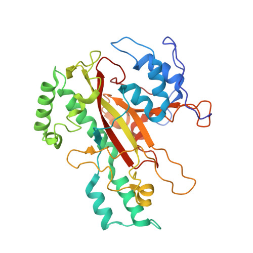
    1IU4

  • PubMed Abstract: 

    The crystal structure of a microbial transglutaminase from Streptoverticillium mobaraense has been determined at 2.4 A resolution. The protein folds into a plate-like shape, and has one deep cleft at the edge of the molecule. Its overall structure is completely different from that of the factor XIII-like transglutaminase, which possesses a cysteine protease-like catalytic triad. The catalytic residue, Cys(64), exists at the bottom of the cleft. Asp(255) resides at the position nearest to Cys(64) and is also adjacent to His(274). Interestingly, Cys(64), Asp(255), and His(274) superimpose well on the catalytic triad "Cys-His-Asp" of the factor XIII-like transglutaminase, in this order. The secondary structure frameworks around these residues are also similar to each other. These results imply that both transglutaminases are related by convergent evolution; however, the microbial transglutaminase has developed a novel catalytic mechanism specialized for the cross-linking reaction. The structure accounts well for the catalytic mechanism, in which Asp(255) is considered to be enzymatically essential, as well as for the causes of the higher reaction rate, the broader substrate specificity, and the lower deamidation activity of this enzyme.


  • Organizational Affiliation
    • Central Research Laboratories, Ajinomoto Company Inc., 1-1 Suzuki-cho, Kawasaki-ku, Kanagawa 210-8681, Japan.

Macromolecule Content 

  • Total Structure Weight: 151.67 kDa 
  • Atom Count: 11,262 
  • Modeled Residue Count: 1,324 
  • Deposited Residue Count: 1,324 
  • Unique protein chains: 1

Macromolecules

Find similar proteins by:|  3D Structure
Entity ID: 1
MoleculeChains  Sequence LengthOrganismDetailsImage
microbial transglutaminase
A, B, C, D
331Streptomyces mobaraensisMutation(s): 0 
EC: 2.3.2.13
UniProt
Find proteins for P81453 (Streptomyces mobaraensis)
Explore P81453 
Go to UniProtKB:  P81453
Entity Groups
Sequence Clusters30% Identity50% Identity70% Identity90% Identity95% Identity100% Identity
UniProt GroupP81453
Sequence Annotations
Expand
Reference Sequence

Experimental Data & Validation

Experimental Data

  • Method: X-RAY DIFFRACTION
  • Resolution: 2.40 Å
  • R-Value Free:  0.266 (Depositor) 
  • R-Value Work:  0.199 (Depositor), 0.207 (DCC) 
  • R-Value Observed: 0.203 (Depositor) 
Space Group: P 1 21 1
Unit Cell:
Length ( Å )Angle ( ˚ )
a = 78.41α = 90
b = 117.12β = 112.8
c = 85.74γ = 90
Software Package:
Software NamePurpose
DENZOdata reduction
SCALEPACKdata scaling
MLPHAREphasing
X-PLORrefinement

Structure Validation

View Full Validation Report


Entry History 

Deposition Data

Revision History  (Full details and data files)

  • Version 1.0: 2002-08-27
    Type: Initial release
  • Version 1.1: 2008-04-27
    Changes: Version format compliance
  • Version 1.2: 2011-07-13
    Changes: Version format compliance
  • Version 1.3: 2023-12-27
    Changes: Data collection, Database references