1INR | pdb_00001inr

CYTOKINE SYNTHESIS


Experimental Data Snapshot

  • Method: X-RAY DIFFRACTION
  • Resolution: 2.00 Å
  • R-Value Free: 
    0.337 (Depositor) 
  • R-Value Work: 
    0.221 (Depositor) 
  • R-Value Observed: 
    0.221 (Depositor) 

wwPDB Validation 3D Report Full Report

Validation slider image for 1INR

This is version 1.3 of the entry. See complete history

Literature

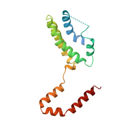
Crystal structure of interleukin 10 reveals an interferon gamma-like fold.

Walter, M.R.Nagabhushan, T.L.

(1995) Biochemistry 34: 12118-12125

  • DOI: https://doi.org/10.1021/bi00038a004
  • Primary Citation Related Structures: 
    1INR

  • PubMed Abstract: 

    The crystal structure of recombinant human interleukin 10 (rhIL-10) has been determined by X-ray crystallography at 2.0 A resolution. Interleukin 10 is a dimer composed of identical polypeptide chains related by a 2-fold axis. The molecule is predominantly alpha-helical. The main-chain fold resembles that of interferon gamma (IFN-gamma) in which the structural integrity of each domain is dependent on the intertwining of helices from each peptide chain. Comparison of rhIL-10 and IFN-gamma reveals differences in helix lengths and orientations of the 2-fold related domains. Interleukin 10 and IFN-gamma contain several conserved residues in their internal cores which suggest a possible "fingerprint" for detection of other members of this fold.


  • Organizational Affiliation
    • Department of Pharmacology, University of Alabama at Birmingham 35294, USA.

Macromolecule Content 

  • Total Structure Weight: 18.67 kDa 
  • Atom Count: 1,114 
  • Modeled Residue Count: 132 
  • Deposited Residue Count: 160 
  • Unique protein chains: 1

Macromolecules

Find similar proteins by:|  3D Structure
Entity ID: 1
MoleculeChains  Sequence LengthOrganismDetailsImage
INTERLEUKIN-10160Homo sapiensMutation(s): 0 
UniProt & NIH Common Fund Data Resources
Find proteins for P22301 (Homo sapiens)
Explore P22301 
Go to UniProtKB:  P22301
PHAROS:  P22301
GTEx:  ENSG00000136634 
Entity Groups
Sequence Clusters30% Identity50% Identity70% Identity90% Identity95% Identity100% Identity
UniProt GroupP22301
Sequence Annotations
Expand
Reference Sequence

Experimental Data & Validation

Experimental Data

  • Method: X-RAY DIFFRACTION
  • Resolution: 2.00 Å
  • R-Value Free:  0.337 (Depositor) 
  • R-Value Work:  0.221 (Depositor) 
  • R-Value Observed: 0.221 (Depositor) 
Space Group: P 43 21 2
Unit Cell:
Length ( Å )Angle ( ˚ )
a = 36.37α = 90
b = 36.37β = 90
c = 220.97γ = 90
Software Package:
Software NamePurpose
DENZOdata reduction
SCALEPACKdata scaling
X-PLORmodel building
X-PLORrefinement
X-PLORphasing

Structure Validation

View Full Validation Report


Entry History 

Deposition Data

  • Released Date: 1996-10-14 
  • Deposition Author(s): Walter, M.R.

Revision History  (Full details and data files)

  • Version 1.0: 1996-10-14
    Type: Initial release
  • Version 1.1: 2008-03-24
    Changes: Version format compliance
  • Version 1.2: 2011-07-13
    Changes: Derived calculations, Version format compliance
  • Version 1.3: 2024-10-30
    Changes: Data collection, Database references, Structure summary