Download Mendeley
Structure-function analysis of a phage display-derived peptide that binds to insulin-like growth factor binding protein 1.
Skelton, N.J., Chen, Y.M., Dubree, N., Quan, C., Jackson, D.Y., Cochran, A., Zobel, K., Deshayes, K., Baca, M., Pisabarro, M.T., Lowman, H.B.(2001) Biochemistry 40: 8487-8498
- PubMed: 11456486 Search on PubMed
- DOI: https://doi.org/10.1021/bi0103866
- Primary Citation Related Structures:
1GJE, 1GJF, 1GJG, 1IMW, 1IN2, 1IN3 - PubMed Abstract:
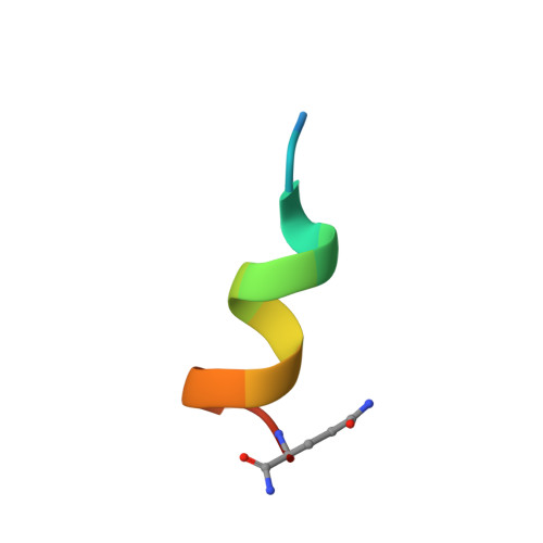
Highly structured, peptide antagonists of the interaction between insulin-like growth factor 1 (IGF-I) and IGF binding protein 1 (IGFBP-1) have recently been discovered by phage display of naïve peptide libraries [Lowman, H. B., et al. (1998) Biochemistry 37, 8870--8878]. We now report a detailed analysis of the features of this turn-helix peptide motif that are necessary for IGFBP-1 binding and structural integrity. Further rounds of phage randomization indicate the importance of residues contributing to a hydrophobic patch on one face of the helix. Alanine-scanning substitutions confirm that the hydrophobic residues are necessary for binding. However, structural analysis by NMR spectroscopy indicates that some of these analogues are less well folded. Structured, high-affinity analogues that lack the disulfide bond were prepared by introducing a covalent constraint between side chains at positions i and i + 7 or i + 8 within the helix. Analogues based on this scaffold demonstrate that a helical conformation is present in the bound state, and that hydrophobic side chains in this helix, and residues immediately preceding it, interact with IGFBP-1. By comparison of alanine scanning data for IGF-I and the turn-helix peptide, we propose a model for common surface features of these molecules that recognize IGFBP-1.
- Department of Protein Engineering, Genentech, Inc., 1 DNA Way, South San Francisco, California 94080, USA. skelly@gene.com
Organizational Affiliation: