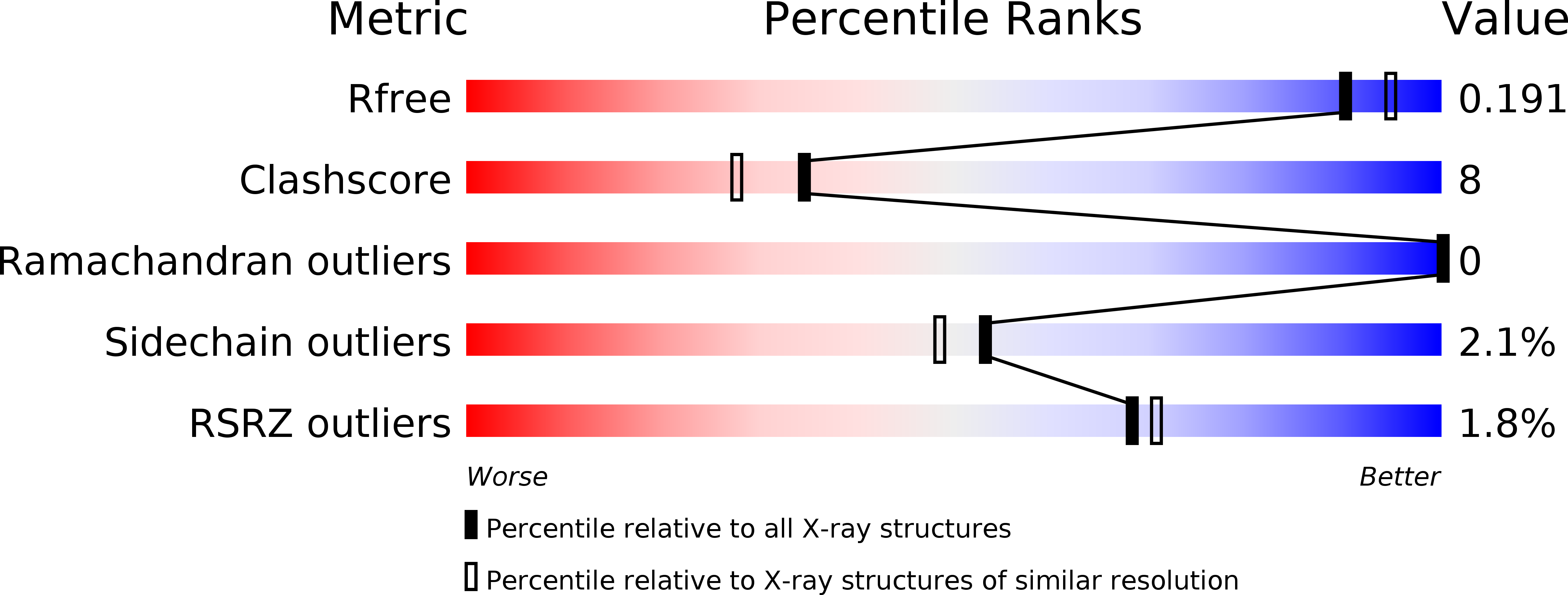
X-ray crystal structure of the trimeric N-terminal domain of gephyrin.
Sola, M., Kneussel, M., Heck, I.S., Betz, H., Weissenhorn, W.(2001) J Biological Chem 276: 25294-25301
- PubMed: 11325967
- DOI: https://doi.org/10.1074/jbc.M101923200
- Primary Citation of Related Structures:
1IHC - PubMed Abstract:
Gephyrin is a ubiquitously expressed protein that, in the central nervous system, forms a submembraneous scaffold for anchoring inhibitory neurotransmitter receptors in the postsynaptic membrane. The N- and C-terminal domains of gephyrin are homologous to the Escherichia coli enzymes MogA and MoeA, respectively, both of which are involved in molybdenum cofactor biosynthesis. This enzymatic pathway is highly conserved from bacteria to mammals, as underlined by the ability of gephyrin to rescue molybdenum cofactor deficiencies in different organisms. Here we report the x-ray crystal structure of the N-terminal domain (amino acids 2-188) of rat gephyrin at 1.9-A resolution. Gephyrin-(2-188) forms trimers in solution, and a sequence motif thought to be involved in molybdopterin binding is highly conserved between gephyrin and the E. coli protein. The atomic structure of gephyrin-(2-188) resembles MogA, albeit with two major differences. The path of the C-terminal ends of gephyrin-(2-188) indicates that the central and C-terminal domains, absent in this structure, should follow a similar 3-fold arrangement as the N-terminal region. In addition, a central beta-hairpin loop found in MogA is lacking in gephyrin-(2-188). Despite these differences, both structures show a high degree of surface charge conservation, which is consistent with their common catalytic function.
Organizational Affiliation:
European Molecular Biology Laboratory, 6 rue Jules Horowitz, B.P.181, 38042 Grenoble Cedex 9, France.


