1IGR | pdb_00001igr

Type 1 Insulin-like growth factor receptor (DOMAINS 1-3)


Experimental Data Snapshot

  • Method: X-RAY DIFFRACTION
  • Resolution: 2.60 Å
  • R-Value Free: 
    0.304 (Depositor) 
  • R-Value Work: 
    0.237 (Depositor) 

wwPDB Validation 3D Report Full Report

Validation slider image for 1IGR

This is version 2.2 of the entry. See complete history

Literature

Crystal structure of the first three domains of the type-1 insulin-like growth factor receptor.

Garrett, T.P.McKern, N.M.Lou, M.Frenkel, M.J.Bentley, J.D.Lovrecz, G.O.Elleman, T.C.Cosgrove, L.J.Ward, C.W.

(1998) Nature 394: 395-399

  • DOI: https://doi.org/10.1038/28668
  • Primary Citation Related Structures: 
    1IGR

  • PubMed Abstract: 

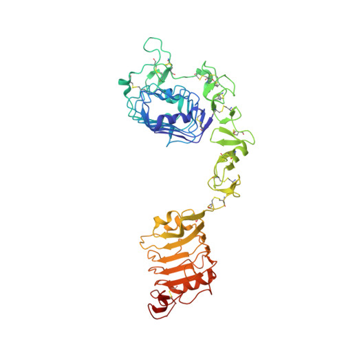
    The type-1 insulin-like growth-factor receptor (IGF-1R) and insulin receptor (IR) are closely related members of the tyrosine-kinase receptor superfamily. IR is essential for glucose homeostasis, whereas IGF-1R is involved in both normal growth and development and malignant transformation. Homologues of these receptors are found in animals as simple as cnidarians. The epidermal growth-factor receptor (EGFR) family is closely related to the IR family and has significant sequence identity to the extracellular portion we describe here. We now present the structure of the first three domains of IGF-IR (L1-Cys-rich-L2) determined to 2.6 A resolution. The L domains each consist of a single-stranded right-handed beta-helix. The Cys-rich region is composed of eight disulphide-bonded modules, seven of which form a rod-shaped domain with modules associated in an unusual manner. The three domains surround a central space of sufficient size to accommodate a ligand molecule. Although the fragment (residues 1-462) does not bind ligand, many of the determinants responsible for hormone binding and ligand specificity map to this central site. This structure therefore shows how the IR subfamily might interact with their ligands.


  • Organizational Affiliation
    • Biomolecular Research Institute, Parkville, Victoria, Australia. tom.barrett@bioresi.com.au

Macromolecule Content 

  • Total Structure Weight: 57.07 kDa 
  • Atom Count: 3,900 
  • Modeled Residue Count: 471 
  • Deposited Residue Count: 478 
  • Unique protein chains: 1

Macromolecules

Find similar proteins by:|  3D Structure
Entity ID: 1
MoleculeChains  Sequence LengthOrganismDetailsImage
INSULIN-LIKE GROWTH FACTOR RECEPTOR 1478Homo sapiensMutation(s): 0 
EC: 2.7.10.1
UniProt & NIH Common Fund Data Resources
Find proteins for P08069 (Homo sapiens)
Explore P08069 
Go to UniProtKB:  P08069
PHAROS:  P08069
GTEx:  ENSG00000140443 
Entity Groups
Sequence Clusters30% Identity50% Identity70% Identity90% Identity95% Identity100% Identity
UniProt GroupP08069
Glycosylation
Glycosylation Sites: 4Go to GlyGen: P08069-1
Sequence Annotations
Expand
Reference Sequence

Oligosaccharides

Help  
Entity ID: 2
MoleculeChains Length2D Diagram GlycosylationD Interactions
2-acetamido-2-deoxy-beta-D-glucopyranose-(1-4)-[alpha-L-fucopyranose-(1-6)]2-acetamido-2-deoxy-beta-D-glucopyranose
B
3N-Glycosylation
Glycosylation Resources
GlyTouCan: G21290RB
GlyCosmos: G21290RB
GlyGen: G21290RB
Entity ID: 3
MoleculeChains Length2D Diagram GlycosylationD Interactions
alpha-D-mannopyranose-(1-3)-beta-D-mannopyranose-(1-4)-2-acetamido-2-deoxy-beta-D-glucopyranose-(1-4)-[alpha-L-fucopyranose-(1-6)]2-acetamido-2-deoxy-beta-D-glucopyranose
C
5N-Glycosylation
Glycosylation Resources
GlyTouCan: G42466VF
GlyCosmos: G42466VF
GlyGen: G42466VF

Small Molecules

Ligands 2 Unique
IDChains Name / Formula / InChI Key2D Diagram3D Interactions
NAG

Query on NAG


Download:Ideal Coordinates CCD File
D [auth A],
E [auth A]
2-acetamido-2-deoxy-beta-D-glucopyranose
C8 H15 N O6
OVRNDRQMDRJTHS-FMDGEEDCSA-N
SO4

Query on SO4


Download:Ideal Coordinates CCD File
F [auth A]
G [auth A]
H [auth A]
I [auth A]
J [auth A]
F [auth A],
G [auth A],
H [auth A],
I [auth A],
J [auth A],
K [auth A],
L [auth A],
M [auth A]
SULFATE ION
O4 S
QAOWNCQODCNURD-UHFFFAOYSA-L

Experimental Data & Validation

Experimental Data

  • Method: X-RAY DIFFRACTION
  • Resolution: 2.60 Å
  • R-Value Free:  0.304 (Depositor) 
  • R-Value Work:  0.237 (Depositor) 
Space Group: P 21 21 21
Unit Cell:
Length ( Å )Angle ( ˚ )
a = 77.39α = 90
b = 99.72β = 90
c = 120.29γ = 90
Software Package:
Software NamePurpose
DENZOdata reduction
SCALEPACKdata scaling
PROTEINmodel building
MLPHAREphasing
DMmodel building
REFMACrefinement
PROTEINphasing
DMphasing

Structure Validation

View Full Validation Report


Entry History 

Deposition Data

Revision History  (Full details and data files)

  • Version 1.0: 1999-09-27
    Type: Initial release
  • Version 1.1: 2008-04-27
    Changes: Version format compliance
  • Version 1.2: 2011-07-13
    Changes: Non-polymer description, Version format compliance
  • Version 1.3: 2018-04-04
    Changes: Data collection
  • Version 2.0: 2020-07-29
    Type: Remediation
    Reason: Carbohydrate remediation
    Changes: Advisory, Atomic model, Data collection, Derived calculations, Structure summary
  • Version 2.1: 2023-12-27
    Changes: Data collection, Database references, Structure summary
  • Version 2.2: 2024-10-30
    Changes: Structure summary