1IGO | pdb_00001igo

Family 11 xylanase


Experimental Data Snapshot

  • Method: X-RAY DIFFRACTION
  • Resolution: 2.20 Å
  • R-Value Free: 
    0.272 (Depositor), 0.270 (DCC) 
  • R-Value Work: 
    0.208 (Depositor), 0.207 (DCC) 
  • R-Value Observed: 
    0.208 (Depositor) 

Starting Model: experimental
View more details

wwPDB Validation 3D Report Full Report

Validation slider image for 1IGO

This is version 1.3 of the entry. See complete history

Literature

Characterization of a family 11 xylanase from Bacillus subtillis B230 used for paper bleaching.

Oakley, A.J.Heinrich, T.Thompson, C.A.Wilce, M.C.

(2003) Acta Crystallogr D Biol Crystallogr 59: 627-636

  • DOI: https://doi.org/10.1107/s0907444903001227
  • Primary Citation Related Structures: 
    1IGO

  • PubMed Abstract: 

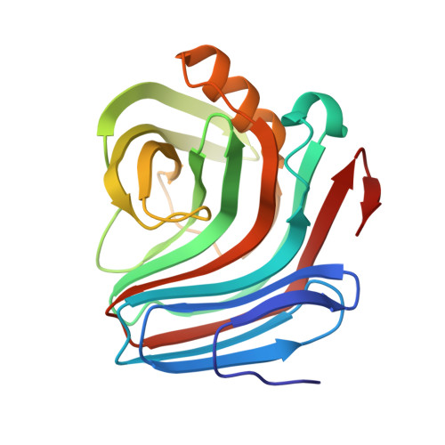
    Enzymes such as family 11 xylanases are increasingly being used for industrial applications. Here, the cloning, structure determination and temperature-stability data of a family 11 xylanase, Xyn11X, from the alkali-tolerant Bacillus subtilis subspecies B230 are reported. This enzyme, which degrades xylan polymers, is being produced on an industrial scale for use in the paper-bleaching industry. Xyn11X adopts the canonical family 11 xylanase fold. It has a greater abundance of side chain to side chain hydrogen bonds compared with all other family 11 xylanase crystal structures. Means by which the thermostability of Xyn11X might be improved are suggested.


  • Organizational Affiliation
    • School of Biomedical and Chemical Sciences, School of Medicine and Pharmacology, University of Western Australia, Nedlands, Australia.

Macromolecule Content 

  • Total Structure Weight: 45.26 kDa 
  • Atom Count: 3,300 
  • Modeled Residue Count: 410 
  • Deposited Residue Count: 410 
  • Unique protein chains: 1

Macromolecules

Find similar proteins by:|  3D Structure
Entity ID: 1
MoleculeChains  Sequence LengthOrganismDetailsImage
family 11 xylanase
A, B
205Bacillus subtilisMutation(s): 0 
EC: 3.2.1.8
UniProt
Find proteins for Q7SID8 (Bacillus subtilis)
Explore Q7SID8 
Go to UniProtKB:  Q7SID8
Entity Groups
Sequence Clusters30% Identity50% Identity70% Identity90% Identity95% Identity100% Identity
UniProt GroupQ7SID8
Sequence Annotations
Expand
Reference Sequence

Small Molecules

Ligands 1 Unique
IDChains Name / Formula / InChI Key2D Diagram3D Interactions
SO4

Query on SO4


Download:Ideal Coordinates CCD File
C [auth B]SULFATE ION
O4 S
QAOWNCQODCNURD-UHFFFAOYSA-L

Experimental Data & Validation

Experimental Data

  • Method: X-RAY DIFFRACTION
  • Resolution: 2.20 Å
  • R-Value Free:  0.272 (Depositor), 0.270 (DCC) 
  • R-Value Work:  0.208 (Depositor), 0.207 (DCC) 
  • R-Value Observed: 0.208 (Depositor) 
Space Group: P 1 21 1
Unit Cell:
Length ( Å )Angle ( ˚ )
a = 49.19α = 90
b = 52.54β = 97.83
c = 65.41γ = 90
Software Package:
Software NamePurpose
DENZOdata reduction
SCALEPACKdata scaling
MOLREPphasing
CNSrefinement

Structure Validation

View Full Validation Report


Entry History 

Deposition Data

Revision History  (Full details and data files)

  • Version 1.0: 2002-04-18
    Type: Initial release
  • Version 1.1: 2008-04-27
    Changes: Version format compliance
  • Version 1.2: 2011-07-13
    Changes: Version format compliance
  • Version 1.3: 2023-08-16
    Changes: Data collection, Database references, Derived calculations, Refinement description