1IFV | pdb_00001ifv

CRYSTAL STRUCTURE OF PATHOGENESIS-RELATED PROTEIN LLPR10.1B FROM YELLOW LUPINE


Experimental Data Snapshot

  • Method: X-RAY DIFFRACTION
  • Resolution: 2.25 Å
  • R-Value Free: 
    0.239 (Depositor), 0.219 (DCC) 
  • R-Value Work: 
    0.186 (Depositor), 0.185 (DCC) 
  • R-Value Observed: 
    0.188 (Depositor) 

Starting Model: experimental
View more details

wwPDB Validation 3D Report Full Report

Validation slider image for 1IFV

This is version 1.4 of the entry. See complete history

Literature

Crystal structures of two homologous pathogenesis-related proteins from yellow lupine.

Biesiadka, J.Bujacz, G.Sikorski, M.M.Jaskolski, M.

(2002) J Mol Biology 319: 1223-1234

  • DOI: https://doi.org/10.1016/S0022-2836(02)00385-6
  • Primary Citation Related Structures: 
    1ICX, 1IFV

  • PubMed Abstract: 

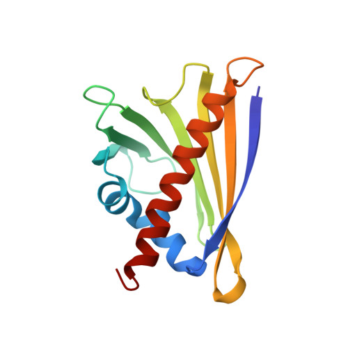
    Pathogenesis-related class 10 (PR10) proteins are restricted to the plant kingdom where they are coded by multigene families and occur at high levels. In spite of their abundance, their physiological role is obscure although members of a distantly related subclass (cytokinin-specific binding proteins) are known to bind plant hormones. PR10 proteins are of special significance in legume plants where their expression patterns are related to infection by the symbiotic, nitrogen-fixing bacteria. Here we present the first crystal structures of classic PR10 proteins representing two homologues from one subclass in yellow lupine. The general fold is similar and, as in a birch pollen allergen, consists of a seven-stranded beta-sheet wrapped around a long C-terminal helix. The mouth of a large pocket formed between the beta-sheet and the helix seems a likely site for ligand binding. The shape of the pocket varies because, in variance with the rigid beta-sheet, the helix shows unusual conformational variability consisting in bending, disorder, and axial shifting. A surface loop, proximal to the entrance to the internal cavity, shows an unusual structural conservation and rigidity in contrast to the high glycine content in its sequence. The loop is different from the so-called glycine-rich P-loops that bind phosphate groups of nucleotides, but it is very likely that it does play a role in ligand binding in PR10 proteins.


  • Organizational Affiliation
    • Institute of Bioorganic Chemistry, Polish Academy of Sciences, 61-704 Poznan, Poland.

Macromolecule Content 

  • Total Structure Weight: 33.09 kDa 
  • Atom Count: 2,448 
  • Modeled Residue Count: 310 
  • Deposited Residue Count: 310 
  • Unique protein chains: 1

Macromolecules

Find similar proteins by:|  3D Structure
Entity ID: 1
MoleculeChains  Sequence LengthOrganismDetailsImage
PROTEIN LLR18B
A, B
155Lupinus luteusMutation(s): 0 
EC: 3.1.27
UniProt
Find proteins for P52779 (Lupinus luteus)
Explore P52779 
Go to UniProtKB:  P52779
Entity Groups
Sequence Clusters30% Identity50% Identity70% Identity90% Identity95% Identity100% Identity
UniProt GroupP52779
Sequence Annotations
Expand
Reference Sequence

Experimental Data & Validation

Experimental Data

  • Method: X-RAY DIFFRACTION
  • Resolution: 2.25 Å
  • R-Value Free:  0.239 (Depositor), 0.219 (DCC) 
  • R-Value Work:  0.186 (Depositor), 0.185 (DCC) 
  • R-Value Observed: 0.188 (Depositor) 
Space Group: P 21 21 2
Unit Cell:
Length ( Å )Angle ( ˚ )
a = 64.88α = 90
b = 107.445β = 90
c = 46.879γ = 90
Software Package:
Software NamePurpose
EPMRphasing
CNSrefinement
DENZOdata reduction
SCALEPACKdata scaling

Structure Validation

View Full Validation Report


Entry History 

Deposition Data

Revision History  (Full details and data files)

  • Version 1.0: 2002-07-10
    Type: Initial release
  • Version 1.1: 2008-04-27
    Changes: Version format compliance
  • Version 1.2: 2011-07-13
    Changes: Version format compliance
  • Version 1.3: 2024-02-07
    Changes: Data collection, Database references
  • Version 1.4: 2024-04-03
    Changes: Refinement description