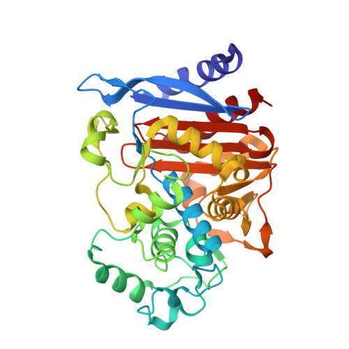
Structures of ceftazidime and its transition-state analogue in complex with AmpC beta-lactamase: implications for resistance mutations and inhibitor design.
Powers, R.A., Caselli, E., Focia, P.J., Prati, F., Shoichet, B.K.(2001) Biochemistry 40: 9207-9214
- PubMed: 11478888 Search on PubMed
- DOI: https://doi.org/10.1021/bi0109358
- Primary Citation Related Structures:
1IEL, 1IEM - PubMed Abstract:
Third-generation cephalosporins are widely used beta-lactam antibiotics that resist hydrolysis by beta-lactamases. Recently, mutant beta-lactamases that rapidly inactivate these drugs have emerged. To investigate why third-generation cephalosporins are relatively stable to wild-type class C beta-lactamases and how mutant enzymes might overcome this, the structures of the class C beta-lactamase AmpC in complex with the third-generation cephalosporin ceftazidime and with a transition-state analogue of ceftazidime were determined by X-ray crystallography to 2.0 and 2.3 A resolution, respectively. Comparison of the acyl-enzyme structures of ceftazidime and loracarbef, a beta-lactam substrate, reveals that the conformation of ceftazidime in the active site differs from that of substrates. Comparison of the structures of the acyl-enzyme intermediate and the transition-state analogue suggests that ceftazidime blocks formation of the tetrahedral transition state, explaining why it is an inhibitor of AmpC. Ceftazidime cannot adopt a conformation competent for catalysis due to steric clashes that would occur with conserved residues Val211 and Tyr221. The X-ray crystal structure of the mutant beta-lactamase GC1, which has improved activity against third-generation cephalosporins, suggests that a tandem tripeptide insertion in the Omega loop, which contains Val211, has caused a shift of this residue and also of Tyr221 that would allow ceftazidime and other third-generation cephalosporins to adopt a more catalytically competent conformation. These structural differences may explain the extended spectrum activity of GC1 against this class of cephalosporins. In addition, the complexed structure of the transition-state analogue inhibitor (K(i) 20 nM) with AmpC reveals potential opportunities for further inhibitor design.
- Department of Molecular Pharmacology and Biological Chemistry, Northwestern University, 303 East Chicago Avenue, Chicago, Illinois 60611, USA.
Organizational Affiliation: