1ID0 | pdb_00001id0

CRYSTAL STRUCTURE OF THE NUCLEOTIDE BOND CONFORMATION OF PHOQ KINASE DOMAIN


Experimental Data Snapshot

  • Method: X-RAY DIFFRACTION
  • Resolution: 1.60 Å
  • R-Value Free: 
    0.233 (Depositor), 0.255 (DCC) 
  • R-Value Work: 
    0.199 (Depositor), 0.219 (DCC) 

wwPDB Validation 3D Report Full Report

Validation slider image for 1ID0

Ligand Structure Quality Assessment 


This is version 1.4 of the entry. See complete history

Literature

Structural and mutational analysis of the PhoQ histidine kinase catalytic domain. Insight into the reaction mechanism.

Marina, A.Mott, C.Auyzenberg, A.Hendrickson, W.A.Waldburger, C.D.

(2001) J Biological Chem 276: 41182-41190

  • DOI: https://doi.org/10.1074/jbc.M106080200
  • Primary Citation Related Structures: 
    1ID0

  • PubMed Abstract: 

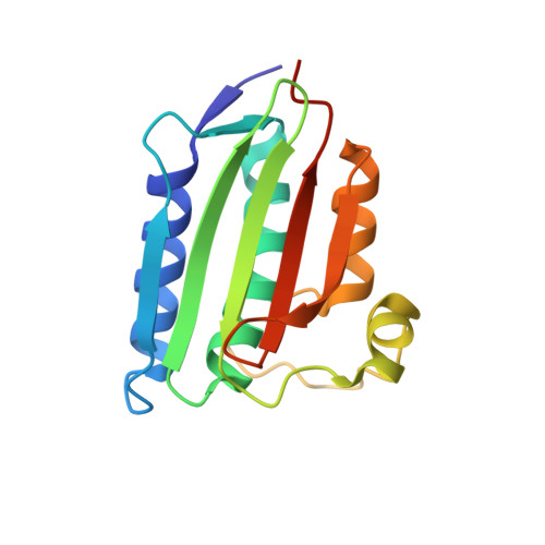
    PhoQ is a transmembrane histidine kinase belonging to the family of two-component signal transducing systems common in prokaryotes and lower eukaryotes. In response to changes in environmental Mg(2+) concentration, PhoQ regulates the level of phosphorylated PhoP, its cognate transcriptional response-regulator. The PhoQ cytoplasmic region comprises two independently folding domains: the histidine-containing phosphotransfer domain and the ATP-binding kinase domain. We have determined the structure of the kinase domain of Escherichia coli PhoQ complexed with the non-hydrolyzable ATP analog adenosine 5'-(beta,gamma-imino)triphosphate and Mg(2+). Nucleotide binding appears to be accompanied by conformational changes in the loop that surrounds the ATP analog (ATP-lid) and has implications for interactions with the substrate phosphotransfer domain. The high resolution (1.6 A) structure reveals a detailed view of the nucleotide-binding site, allowing us to identify potential catalytic residues. Mutagenic analyses of these residues provide new insights into the catalytic mechanism of histidine phosphorylation in the histidine kinase family. Comparison with the active site of the related GHL ATPase family reveals differences that are proposed to account for the distinct functions of these proteins.


  • Organizational Affiliation
    • Department of Biochemistry and Molecular Biophysics, Columbia University, New York, New York 10032, USA.

Macromolecule Content 

  • Total Structure Weight: 17.38 kDa 
  • Atom Count: 1,447 
  • Modeled Residue Count: 146 
  • Deposited Residue Count: 152 
  • Unique protein chains: 1

Macromolecules

Find similar proteins by:|  3D Structure
Entity ID: 1
MoleculeChains  Sequence LengthOrganismDetailsImage
PHOQ HISTIDINE KINASE152Escherichia coliMutation(s): 0 
EC: 2.7.1.37 (PDB Primary Data), 2.7.13.3 (UniProt), 3.1.3 (UniProt)
UniProt
Find proteins for P23837 (Escherichia coli (strain K12))
Explore P23837 
Go to UniProtKB:  P23837
Entity Groups
Sequence Clusters30% Identity50% Identity70% Identity90% Identity95% Identity100% Identity
UniProt GroupP23837
Sequence Annotations
Expand
Reference Sequence

Small Molecules

Ligands 2 Unique
IDChains Name / Formula / InChI Key2D Diagram3D Interactions
ANP

Query on ANP


Download:Ideal Coordinates CCD File
C [auth A]PHOSPHOAMINOPHOSPHONIC ACID-ADENYLATE ESTER
C10 H17 N6 O12 P3
PVKSNHVPLWYQGJ-KQYNXXCUSA-N
MG

Query on MG


Download:Ideal Coordinates CCD File
B [auth A]MAGNESIUM ION
Mg
JLVVSXFLKOJNIY-UHFFFAOYSA-N

Experimental Data & Validation

Experimental Data

  • Method: X-RAY DIFFRACTION
  • Resolution: 1.60 Å
  • R-Value Free:  0.233 (Depositor), 0.255 (DCC) 
  • R-Value Work:  0.199 (Depositor), 0.219 (DCC) 
Space Group: P 21 21 21
Unit Cell:
Length ( Å )Angle ( ˚ )
a = 43.503α = 90
b = 45.004β = 90
c = 71.764γ = 90
Software Package:
Software NamePurpose
MADSYSphasing
MLPHAREphasing
DMmodel building
CNSrefinement
DENZOdata reduction
SCALEPACKdata scaling
DMphasing

Structure Validation

View Full Validation Report


Ligand Structure Quality Assessment 


Entry History 

Deposition Data

Revision History  (Full details and data files)

  • Version 1.0: 2001-10-17
    Type: Initial release
  • Version 1.1: 2008-04-27
    Changes: Version format compliance
  • Version 1.2: 2011-07-13
    Changes: Version format compliance
  • Version 1.3: 2017-02-01
    Changes: Structure summary
  • Version 1.4: 2024-02-07
    Changes: Data collection, Database references, Derived calculations