1IAU

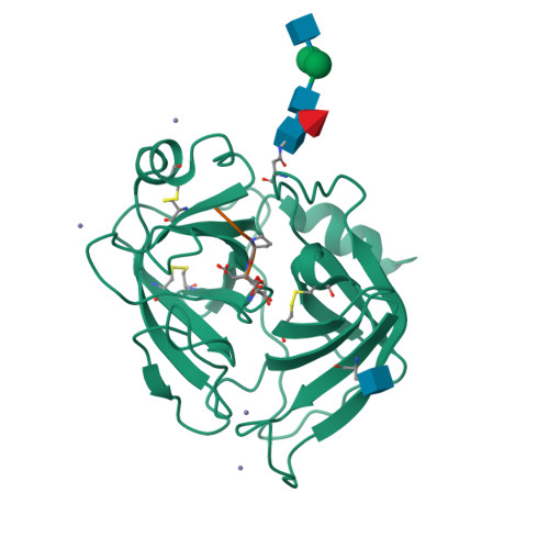
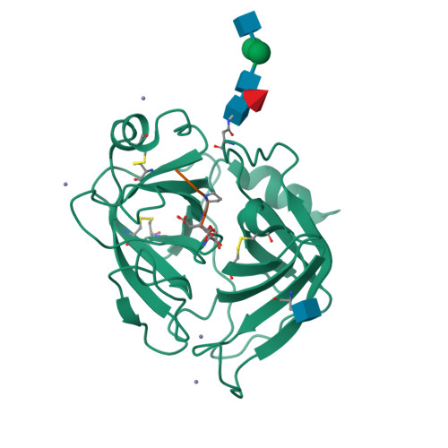
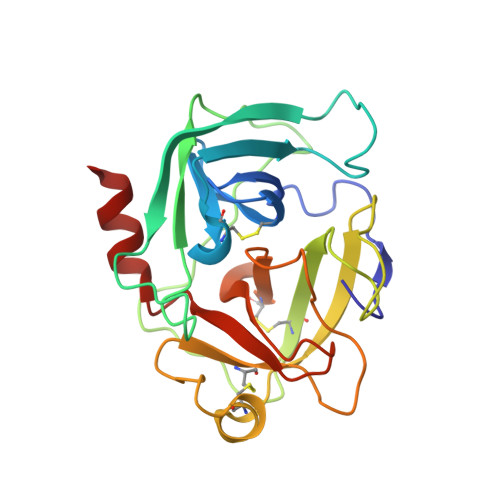
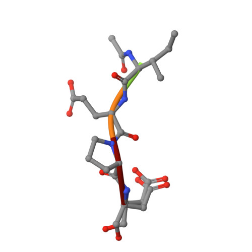
HUMAN GRANZYME B IN COMPLEX WITH AC-IEPD-CHO


Experimental Data Snapshot

  • Method: X-RAY DIFFRACTION
  • Resolution: 2.00 Å
  • R-Value Free: 0.296 
  • R-Value Work: 0.239 

Starting Model: experimental
View more details

wwPDB Validation   3D Report Full Report


Ligand Structure Quality Assessment 


This is version 2.2 of the entry. See complete history


Literature

The three-dimensional structure of human granzyme B compared to caspase-3, key mediators of cell death with cleavage specificity for aspartic acid in P1.

Rotonda, J.Garcia-Calvo, M.Bull, H.G.Geissler, W.M.McKeever, B.M.Willoughby, C.A.Thornberry, N.A.Becker, J.W.

(2001) Chem Biol 8: 357-368

  • DOI: https://doi.org/10.1016/s1074-5521(01)00018-7
  • Primary Citation of Related Structures:  
    1IAU

  • PubMed Abstract: 

    Granzyme B, one of the most abundant granzymes in cytotoxic T-lymphocyte (CTL) granules, and members of the caspase (cysteine aspartyl proteinases) family have a unique cleavage specificity for aspartic acid in P1 and play critical roles in the biochemical events that culminate in cell death. We have determined the three-dimensional structure of the complex of the human granzyme B with a potent tetrapeptide aldehyde inhibitor. The Asp-specific S1 subsite of human granzyme B is significantly larger and less charged than the corresponding Asp-specific site in the apoptosis-promoting caspases, and also larger than the corresponding subsite in rat granzyme B. The above differences account for the variation in substrate specificity among granzyme B, other serine proteases and the caspases, and enable the design of specific inhibitors that can probe the physiological functions of these proteins and the disease states with which they are associated.


  • Organizational Affiliation

    Department of Endocrinology and Chemical Biology, Merck Research Laboratories, Rahway, NJ 07065-0900, USA. rotonda@merck.com


Macromolecules
Find similar proteins by:  (by identity cutoff)  |  3D Structure
Entity ID: 1
MoleculeChains Sequence LengthOrganismDetailsImage
GRANZYME B227Homo sapiensMutation(s): 1 
EC: 3.4.21.79
UniProt & NIH Common Fund Data Resources
Find proteins for P10144 (Homo sapiens)
Explore P10144 
Go to UniProtKB:  P10144
PHAROS:  P10144
GTEx:  ENSG00000100453 
Entity Groups  
Sequence Clusters30% Identity50% Identity70% Identity90% Identity95% Identity100% Identity
UniProt GroupP10144
Glycosylation
Glycosylation Sites: 2Go to GlyGen: P10144-1
Sequence Annotations
Expand
  • Reference Sequence

Find similar proteins by:  Sequence   |   3D Structure  

Entity ID: 2
MoleculeChains Sequence LengthOrganismDetailsImage
acetyl-isoleucyl-glutamyl-prolyl-aspartyl-aldehyde peptide INHIBITOR5N/AMutation(s): 0 
Sequence Annotations
Expand
  • Reference Sequence
Oligosaccharides

Help

Entity ID: 3
MoleculeChains Length2D Diagram Glycosylation3D Interactions
2-acetamido-2-deoxy-beta-D-glucopyranose-(1-4)-[beta-D-mannopyranose-(1-6)]alpha-D-mannopyranose-(1-4)-2-acetamido-2-deoxy-beta-D-glucopyranose-(1-4)-[alpha-L-fucopyranose-(1-6)]2-acetamido-2-deoxy-beta-D-glucopyranose
C
6N-Glycosylation
Glycosylation Resources
GlyTouCan:  G22026YZ
GlyCosmos:  G22026YZ
GlyGen:  G22026YZ
Experimental Data & Validation

Experimental Data

  • Method: X-RAY DIFFRACTION
  • Resolution: 2.00 Å
  • R-Value Free: 0.296 
  • R-Value Work: 0.239 
  • Space Group: P 21 21 21
Unit Cell:
Length ( Å )Angle ( ˚ )
a = 48.873α = 90
b = 75.081β = 90
c = 80.261γ = 90
Software Package:
Software NamePurpose
HKL-2000data collection
SCALEPACKdata scaling
CNXrefinement
HKL-2000data reduction
CNXphasing

Structure Validation

View Full Validation Report


Ligand Structure Quality Assessment 


Entry History 

Deposition Data

Revision History  (Full details and data files)

  • Version 1.0: 2001-05-02
    Type: Initial release
  • Version 1.1: 2008-04-27
    Changes: Version format compliance
  • Version 1.2: 2011-07-13
    Changes: Atomic model, Database references, Derived calculations, Non-polymer description, Structure summary, Version format compliance
  • Version 1.3: 2012-04-04
    Changes: Structure summary
  • Version 1.4: 2012-12-12
    Changes: Other
  • Version 2.0: 2020-07-29
    Type: Remediation
    Reason: Carbohydrate remediation
    Changes: Atomic model, Data collection, Database references, Derived calculations, Structure summary
  • Version 2.1: 2023-08-09
    Changes: Data collection, Database references, Refinement description, Structure summary
  • Version 2.2: 2023-11-15
    Changes: Data collection