1IAK

HISTOCOMPATIBILITY ANTIGEN I-AK


Experimental Data Snapshot

  • Method: X-RAY DIFFRACTION
  • Resolution: 1.90 Å
  • R-Value Free: 0.263 
  • R-Value Work: 0.214 
  • R-Value Observed: 0.214 

Starting Model: experimental
View more details

wwPDB Validation   3D Report Full Report


This is version 1.6 of the entry. See complete history


Literature

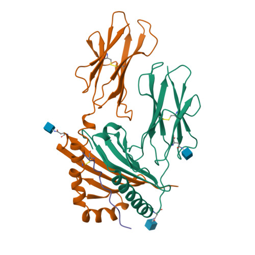
Crystal structure of I-Ak in complex with a dominant epitope of lysozyme.

Fremont, D.H.Monnaie, D.Nelson, C.A.Hendrickson, W.A.Unanue, E.R.

(1998) Immunity 8: 305-317

  • DOI: https://doi.org/10.1016/s1074-7613(00)80536-1
  • Primary Citation of Related Structures:  
    1IAK

  • PubMed Abstract: 

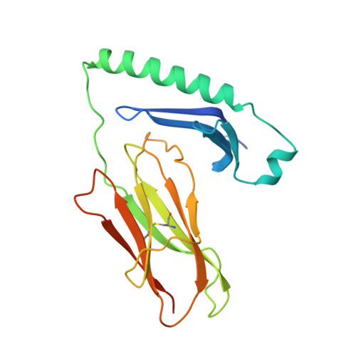
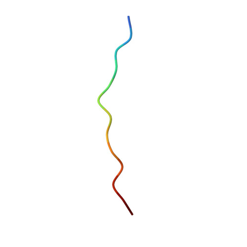
    We have determined the structure of murine MHC class II I-Ak in complex with a naturally processed peptide from hen egg lysozyme (HEL residues 50-62) at 1.9 A resolution. These results provide a structural basis for the I-Ak peptide-binding motif. Binding is established by the deep burial of five anchor side chains into specific pockets of the I-Ak binding groove, with a zen-like fit of an aspartic acid in the P1 pocket. We also show that in the I-Ak alpha chain, a bulge occurs in the first strand of the peptide-binding platform, an insertion probably common to all I-A and HLA-DQ alleles. The I-Ak beta chain has a deletion in the helical region adjacent to the P7 pocket and an insertion in the helical region neighboring the P1 pocket. As a result of these structural features, the extended HEL peptide dips low into the center of the I-Ak groove and reaches toward solvent at its C-terminal end.


  • Organizational Affiliation

    Howard Hughes Medical Institute, Department of Biochemistry and Molecular Biophysics, Columbia University, New York, New York 10032, USA. fremont@immunology.wustl.edu


Macromolecules
Find similar proteins by:  (by identity cutoff)  |  3D Structure
Entity ID: 1
MoleculeChains Sequence LengthOrganismDetailsImage
MHC CLASS II I-AK199Mus musculusMutation(s): 0 
UniProt
Find proteins for P01910 (Mus musculus)
Explore P01910 
Go to UniProtKB:  P01910
Entity Groups  
Sequence Clusters30% Identity50% Identity70% Identity90% Identity95% Identity100% Identity
UniProt GroupP01910
Glycosylation
Glycosylation Sites: 2Go to GlyGen: P01910-1
Sequence Annotations
Expand
  • Reference Sequence
Find similar proteins by:  (by identity cutoff)  |  3D Structure
Entity ID: 2
MoleculeChains Sequence LengthOrganismDetailsImage
MHC CLASS II I-AK185Mus musculusMutation(s): 0 
UniProt
Find proteins for P06343 (Mus musculus)
Explore P06343 
Go to UniProtKB:  P06343
Entity Groups  
Sequence Clusters30% Identity50% Identity70% Identity90% Identity95% Identity100% Identity
UniProt GroupP06343
Glycosylation
Glycosylation Sites: 1
Sequence Annotations
Expand
  • Reference Sequence

Find similar proteins by:  Sequence   |   3D Structure  

Entity ID: 3
MoleculeChains Sequence LengthOrganismDetailsImage
MHC CLASS II I-AKC [auth P]13Mus musculusMutation(s): 0 
UniProt
Find proteins for Q29431 (Ovis aries)
Explore Q29431 
Go to UniProtKB:  Q29431
Entity Groups  
Sequence Clusters30% Identity50% Identity70% Identity90% Identity95% Identity100% Identity
UniProt GroupQ29431
Sequence Annotations
Expand
  • Reference Sequence
Experimental Data & Validation

Experimental Data

  • Method: X-RAY DIFFRACTION
  • Resolution: 1.90 Å
  • R-Value Free: 0.263 
  • R-Value Work: 0.214 
  • R-Value Observed: 0.214 
  • Space Group: P 41 21 2
Unit Cell:
Length ( Å )Angle ( ˚ )
a = 112.25α = 90
b = 112.25β = 90
c = 99.72γ = 90
Software Package:
Software NamePurpose
HKLdata collection
DENZOdata reduction
SCALAdata scaling
Agrovatadata reduction
X-PLORmodel building
X-PLORrefinement
HKLdata reduction
CCP4data scaling
X-PLORphasing

Structure Validation

View Full Validation Report


Entry History 

Deposition Data

Revision History  (Full details and data files)

  • Version 1.0: 1998-04-15
    Type: Initial release
  • Version 1.1: 2008-03-03
    Changes: Version format compliance
  • Version 1.2: 2011-07-13
    Changes: Non-polymer description, Version format compliance
  • Version 1.3: 2011-11-16
    Changes: Atomic model
  • Version 1.4: 2020-07-29
    Type: Remediation
    Reason: Carbohydrate remediation
    Changes: Data collection, Database references, Derived calculations, Refinement description, Structure summary
  • Version 1.5: 2023-08-09
    Changes: Database references, Refinement description, Structure summary
  • Version 1.6: 2024-10-23
    Changes: Data collection, Structure summary