1IA8 | pdb_00001ia8

THE 1.7 A CRYSTAL STRUCTURE OF HUMAN CELL CYCLE CHECKPOINT KINASE CHK1


Experimental Data Snapshot

  • Method: X-RAY DIFFRACTION
  • Resolution: 1.70 Å
  • R-Value Free: 
    0.235 (Depositor) 
  • R-Value Work: 
    0.216 (Depositor) 

Starting Model: experimental
View more details

wwPDB Validation 3D Report Full Report

Validation slider image for 1IA8

This is version 1.5 of the entry. See complete history

Literature

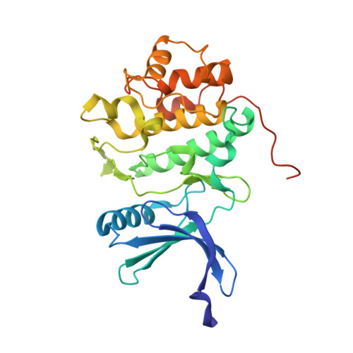
The 1.7 A crystal structure of human cell cycle checkpoint kinase Chk1: implications for Chk1 regulation.

Chen, P.Luo, C.Deng, Y.Ryan, K.Register, J.Margosiak, S.Tempczyk-Russell, A.Nguyen, B.Myers, P.Lundgren, K.Kan, C.C.O'Connor, P.M.

(2000) Cell 100: 681-692

  • DOI: https://doi.org/10.1016/s0092-8674(00)80704-7
  • Primary Citation Related Structures: 
    1IA8

  • PubMed Abstract: 

    The checkpoint kinase Chk1 is an important mediator of cell cycle arrest following DNA damage. The 1.7 A resolution crystal structures of the human Chk1 kinase domain and its binary complex with an ATP analog has revealed an identical open kinase conformation. The secondary structure and side chain interactions stabilize the activation loop of Chk1 and enable kinase activity without phosphorylation of the catalytic domain. Molecular modeling of the interaction of a Cdc25C peptide with Chk1 has uncovered several conserved residues that are important for substrate selectivity. In addition, we found that the less conserved C-terminal region negatively impacts Chk1 kinase activity.


  • Organizational Affiliation
    • Agouron Pharmaceuticals, Inc. San Diego, California 92121, USA. ping.chen@agouron.com

Macromolecule Content 

  • Total Structure Weight: 33.14 kDa 
  • Atom Count: 2,370 
  • Modeled Residue Count: 272 
  • Deposited Residue Count: 289 
  • Unique protein chains: 1

Macromolecules

Find similar proteins by:|  3D Structure
Entity ID: 1
MoleculeChains  Sequence LengthOrganismDetailsImage
CHK1 CHECKPOINT KINASE289Homo sapiensMutation(s): 0 
EC: 2.7.1 (PDB Primary Data), 2.7.11.1 (UniProt)
UniProt & NIH Common Fund Data Resources
Find proteins for O14757 (Homo sapiens)
Explore O14757 
Go to UniProtKB:  O14757
PHAROS:  O14757
GTEx:  ENSG00000149554 
Entity Groups
Sequence Clusters30% Identity50% Identity70% Identity90% Identity95% Identity100% Identity
UniProt GroupO14757
Sequence Annotations
Expand
Reference Sequence

Small Molecules

Ligands 1 Unique
IDChains Name / Formula / InChI Key2D Diagram3D Interactions
SO4

Query on SO4


Download:Ideal Coordinates CCD File
B [auth A]SULFATE ION
O4 S
QAOWNCQODCNURD-UHFFFAOYSA-L

Experimental Data & Validation

Experimental Data

  • Method: X-RAY DIFFRACTION
  • Resolution: 1.70 Å
  • R-Value Free:  0.235 (Depositor) 
  • R-Value Work:  0.216 (Depositor) 
Space Group: P 1 21 1
Unit Cell:
Length ( Å )Angle ( ˚ )
a = 45.2α = 90
b = 65.7β = 93.9
c = 58.1γ = 90
Software Package:
Software NamePurpose
AMoREphasing
PHASESphasing
X-PLORrefinement
DENZOdata reduction
SCALEPACKdata scaling

Structure Validation

View Full Validation Report


Entry History 

Revision History  (Full details and data files)

  • Version 1.0: 2001-04-18
    Type: Initial release
  • Version 1.1: 2008-04-27
    Changes: Version format compliance
  • Version 1.2: 2011-07-13
    Changes: Version format compliance
  • Version 1.3: 2018-01-31
    Changes: Experimental preparation
  • Version 1.4: 2024-02-07
    Changes: Data collection, Database references, Derived calculations
  • Version 1.5: 2024-04-03
    Changes: Refinement description