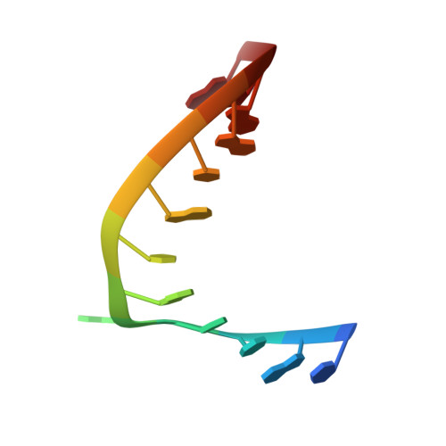
Crystal structure of a model branchpoint-U2 snRNA duplex containing bulged adenosines.
Berglund, J.A., Rosbash, M., Schultz, S.C.(2001) RNA 7: 682-691
- PubMed: 11350032 Search on PubMedSearch on PubMed Central
- DOI: https://doi.org/10.1017/s1355838201002187
- Primary Citation Related Structures:
1I9X - PubMed Abstract:
Bulged nucleotides play a variety of important roles in RNA structure and function, frequently forming tertiary interactions and sometimes even participating in RNA catalysis. In pre-mRNA splicing, the U2 snRNA base pairs with the intron branchpoint sequence (BPS) to form a short RNA duplex that contains a bulged adenosine that ultimately serves as the nucleophile that attacks the 5' splice site. We have determined a 2.18-A resolution crystal structure of a self-complementary RNA designed to mimic the highly conserved yeast (Saccharomyces cerevisiae) branchpoint sequence (5'-UACUAACGUAGUA with the BPS italicized and the branchsite adenosine underlined) base paired with its complementary sequence from U2 snRNA. The structure shows a nearly ideal A-form helix from which two unpaired adenosines flip out. Although the adenosine adjacent to the branchsite adenosine is the one bulged out in the structure described here, either of these adenosines can serve as the nucleophile in mammalian but not in yeast pre-mRNA splicing. In addition, the packing of the bulged RNA helices within the crystal reveals a novel RNA tertiary interaction in which three RNA helices interact through bulged adenosines in the absence of any divalent metal ions.
- Department of Chemistry and Biochemistry, University of Colorado, Boulder 80309, USA. Andy.Berglund@Colorado.edu
Organizational Affiliation: