1I6P | pdb_00001i6p

CRYSTAL STRUCTURE OF E. COLI BETA CARBONIC ANHYDRASE (ECCA)


Experimental Data Snapshot

  • Method: X-RAY DIFFRACTION
  • Resolution: 2.00 Å
  • R-Value Free: 
    0.202 (Depositor) 
  • R-Value Work: 
    0.176 (Depositor) 
  • R-Value Observed: 
    0.176 (Depositor) 

Starting Model: experimental
View more details

wwPDB Validation 3D Report Full Report

Validation slider image for 1I6P

This is version 1.3 of the entry. See complete history

Literature

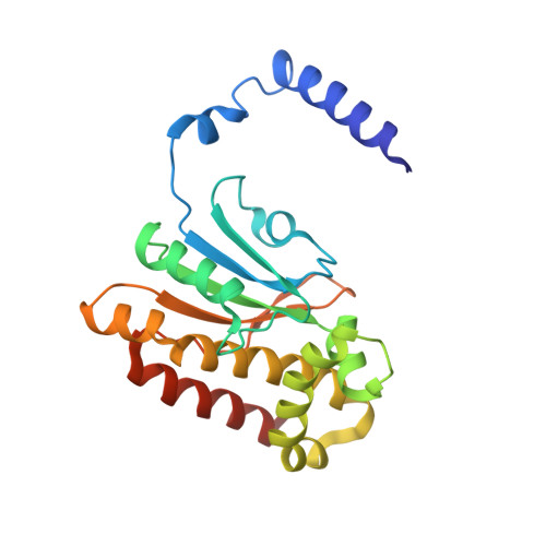
Crystal structure of E. coli beta-carbonic anhydrase, an enzyme with an unusual pH-dependent activity.

Cronk, J.D.Endrizzi, J.A.Cronk, M.R.O'neill, J.W.Zhang, K.Y.

(2001) Protein Sci 10: 911-922

  • DOI: https://doi.org/10.1110/ps.46301
  • Primary Citation Related Structures: 
    1I6O, 1I6P

  • PubMed Abstract: 

    Carbonic anhydrases fall into three distinct evolutionary and structural classes: alpha, beta, and gamma. The beta-class carbonic anhydrases (beta-CAs) are widely distributed among higher plants, simple eukaryotes, eubacteria, and archaea. We have determined the crystal structure of ECCA, a beta-CA from Escherichia coli, to a resolution of 2.0 A. In agreement with the structure of the beta-CA from the chloroplast of the red alga Porphyridium purpureum, the active-site zinc in ECCA is tetrahedrally coordinated by the side chains of four conserved residues. These results confirm the observation of a unique pattern of zinc ligation in at least some beta-CAS: The absence of a water molecule in the inner coordination sphere is inconsistent with known mechanisms of CA activity. ECCA activity is highly pH-dependent in the physiological range, and its expression in yeast complements an oxygen-sensitive phenotype displayed by a beta-CA-deletion strain. The structural and biochemical characterizations of ECCA presented here and the comparisons with other beta-CA structures suggest that ECCA can adopt two distinct conformations displaying widely divergent catalytic rates.


  • Organizational Affiliation
    • Division of Basic Sciences, Fred Hutchinson Cancer Research Center, Seattle, Washington 98109-1024, USA.

Macromolecule Content 

  • Total Structure Weight: 25.2 kDa 
  • Atom Count: 1,778 
  • Modeled Residue Count: 214 
  • Deposited Residue Count: 220 
  • Unique protein chains: 1

Macromolecules

Find similar proteins by:|  3D Structure
Entity ID: 1
MoleculeChains  Sequence LengthOrganismDetailsImage
CARBONIC ANHYDRASE220Escherichia coliMutation(s): 0 
Gene Names: YADF
EC: 4.2.1.1
UniProt
Find proteins for P61517 (Escherichia coli (strain K12))
Explore P61517 
Go to UniProtKB:  P61517
Entity Groups
Sequence Clusters30% Identity50% Identity70% Identity90% Identity95% Identity100% Identity
UniProt GroupP61517
Sequence Annotations
Expand
Reference Sequence

Small Molecules

Ligands 1 Unique
IDChains Name / Formula / InChI Key2D Diagram3D Interactions
ZN

Query on ZN


Download:Ideal Coordinates CCD File
B [auth A]ZINC ION
Zn
PTFCDOFLOPIGGS-UHFFFAOYSA-N

Experimental Data & Validation

Experimental Data

  • Method: X-RAY DIFFRACTION
  • Resolution: 2.00 Å
  • R-Value Free:  0.202 (Depositor) 
  • R-Value Work:  0.176 (Depositor) 
  • R-Value Observed: 0.176 (Depositor) 
Space Group: P 42 21 2
Unit Cell:
Length ( Å )Angle ( ˚ )
a = 68.535α = 90
b = 68.535β = 90
c = 85.877γ = 90
Software Package:
Software NamePurpose
EPMRphasing
CNSrefinement
DENZOdata reduction
SCALEPACKdata scaling

Structure Validation

View Full Validation Report


Entry History 

Deposition Data

Revision History  (Full details and data files)

  • Version 1.0: 2001-05-09
    Type: Initial release
  • Version 1.1: 2008-04-27
    Changes: Version format compliance
  • Version 1.2: 2011-07-13
    Changes: Derived calculations, Version format compliance
  • Version 1.3: 2023-08-09
    Changes: Data collection, Database references, Derived calculations, Refinement description