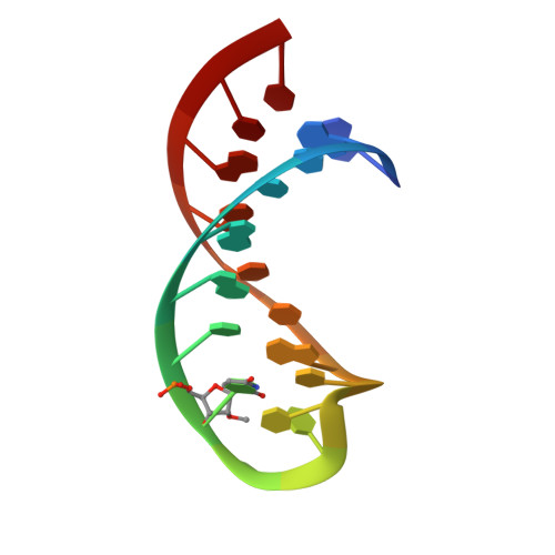
Solution structure of the A loop of 23S ribosomal RNA.
Blanchard, S.C., Puglisi, J.D.(2001) Proc Natl Acad Sci U S A 98: 3720-3725
- PubMed: 11259644 Search on PubMedSearch on PubMed Central
- DOI: https://doi.org/10.1073/pnas.051608498
- Primary Citation Related Structures:
1I3X, 1I3Y - PubMed Abstract:
The A loop is an essential RNA component of the ribosome peptidyltransferase center that directly interacts with aminoacyl (A)-site tRNA. The A loop is highly conserved and contains a ubiquitous 2'-O-methyl ribose modification at position U2552. Here, we present the solution structure of a modified and unmodified A-loop RNA to define both the A-loop fold and the structural impact of the U2552 modification. Solution data reveal that the A-loop RNA has a compact structure that includes a noncanonical base pair between C2556 and U2552. NMR evidence is presented that the N3 position of C2556 has a shifted pKa and that protonation at C2556-N3 changes the C-U pair geometry. Our data indicate that U2552 methylation modifies the A-loop fold, in particular the dynamics and position of residues C2556 and U2555. We compare our structural data with the structure of the A loop observed in a recent 50S crystal structure [Ban, N., Nissen, P., Hansen, J., Moore, P. B. & Steitz, T. A. (2000) Science 289, 905--920; Nissen, P., Hansen, J., Ban, N., Moore, P. B. & Steitz, T. A. (2000) Science 289, 920--930]. The solution and crystal structures of the A loop are dramatically different, suggesting that a structural rearrangement of the A loop must occur on docking into the peptidyltransferase center. Possible roles of this docking event, the shifted pKa of C2556 and the U2552 2'-O-methylation in the mechanism of translation, are discussed.
- Stanford University School of Medicine, Department of Structural Biology, 299 Campus Drive West, Fairchild Building, Stanford, CA 94305-5126, USA.
Organizational Affiliation: