1I0Z | pdb_00001i0z

HUMAN HEART L-LACTATE DEHYDROGENASE H CHAIN, TERNARY COMPLEX WITH NADH AND OXAMATE


Experimental Data Snapshot

  • Method: X-RAY DIFFRACTION
  • Resolution: 2.10 Å
  • R-Value Free: 
    0.230 (Depositor), 0.244 (DCC) 
  • R-Value Work: 
    0.177 (Depositor) 
  • R-Value Observed: 
    0.179 (Depositor) 

Starting Model: experimental
View more details

wwPDB Validation 3D Report Full Report

Validation slider image for 1I0Z

Ligand Structure Quality Assessment 


This is version 1.4 of the entry. See complete history

Literature

Structural basis for altered activity of M- and H-isozyme forms of human lactate dehydrogenase.

Read, J.A.Winter, V.J.Eszes, C.M.Sessions, R.B.Brady, R.L.

(2001) Proteins 43: 175-185

  • DOI: https://doi.org/10.1002/1097-0134(20010501)43:2<175::aid-prot1029>3.0.co;2-#
  • Primary Citation Related Structures: 
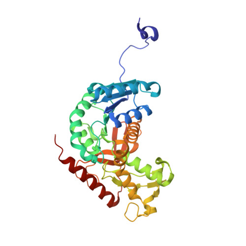
    1I0Z, 1I10

  • PubMed Abstract: 

    Lactate dehydrogenase (LDH) interconverts pyruvate and lactate with concomitant interconversion of NADH and NAD(+). Although crystal structures of a variety of LDH have previously been described, a notable absence has been any of the three known human forms of this glycolytic enzyme. We have now determined the crystal structures of two isoforms of human LDH-the M form, predominantly found in muscle; and the H form, found mainly in cardiac muscle. Both structures have been crystallized as ternary complexes in the presence of the NADH cofactor and oxamate, a substrate-like inhibitor. Although each of these isoforms has different kinetic properties, the domain structure, subunit association, and active-site regions are indistinguishable between the two structures. The pK(a) that governs the K(M) for pyruvate for the two isozymes is found to differ by about 0.94 pH units, consistent with variation in pK(a) of the active-site histidine. The close similarity of these crystal structures suggests the distinctive activity of these enzyme isoforms is likely to result directly from variation of charged surface residues peripheral to the active site, a hypothesis supported by electrostatic calculations based on each structure. Proteins 2001;43:175-185.


  • Organizational Affiliation
    • Department of Biochemistry, University of Bristol, Bristol BS8 1TD, United Kingdom.

Macromolecule Content 

  • Total Structure Weight: 74.61 kDa 
  • Atom Count: 5,522 
  • Modeled Residue Count: 664 
  • Deposited Residue Count: 666 
  • Unique protein chains: 1

Macromolecules

Find similar proteins by:|  3D Structure
Entity ID: 1
MoleculeChains  Sequence LengthOrganismDetailsImage
L-LACTATE DEHYDROGENASE H CHAIN
A, B
333Homo sapiensMutation(s): 0 
Gene Names: LDHB
EC: 1.1.1.27
UniProt & NIH Common Fund Data Resources
Find proteins for P07195 (Homo sapiens)
Explore P07195 
Go to UniProtKB:  P07195
PHAROS:  P07195
GTEx:  ENSG00000111716 
Entity Groups
Sequence Clusters30% Identity50% Identity70% Identity90% Identity95% Identity100% Identity
UniProt GroupP07195
Sequence Annotations
Expand
Reference Sequence

Small Molecules

Binding Affinity Annotations 
IDSourceBinding Affinity
OXM BindingDB:  1I0Z Ki: 9.44e+4 (nM) from 1 assay(s)
IC50: min: 3.38e+4, max: 1.23e+5 (nM) from 3 assay(s)

Experimental Data & Validation

Experimental Data

  • Method: X-RAY DIFFRACTION
  • Resolution: 2.10 Å
  • R-Value Free:  0.230 (Depositor), 0.244 (DCC) 
  • R-Value Work:  0.177 (Depositor) 
  • R-Value Observed: 0.179 (Depositor) 
Space Group: C 2 2 21
Unit Cell:
Length ( Å )Angle ( ˚ )
a = 59.491α = 90
b = 161.683β = 90
c = 137.846γ = 90
Software Package:
Software NamePurpose
DENZOdata reduction
SCALEPACKdata scaling
AMoREphasing
REFMACrefinement

Structure Validation

View Full Validation Report


Ligand Structure Quality Assessment 


Entry History 

Deposition Data

Revision History  (Full details and data files)

  • Version 1.0: 2001-03-28
    Type: Initial release
  • Version 1.1: 2008-04-27
    Changes: Version format compliance
  • Version 1.2: 2011-07-13
    Changes: Derived calculations, Version format compliance
  • Version 1.3: 2014-11-12
    Changes: Structure summary
  • Version 1.4: 2023-08-09
    Changes: Data collection, Database references, Derived calculations, Refinement description